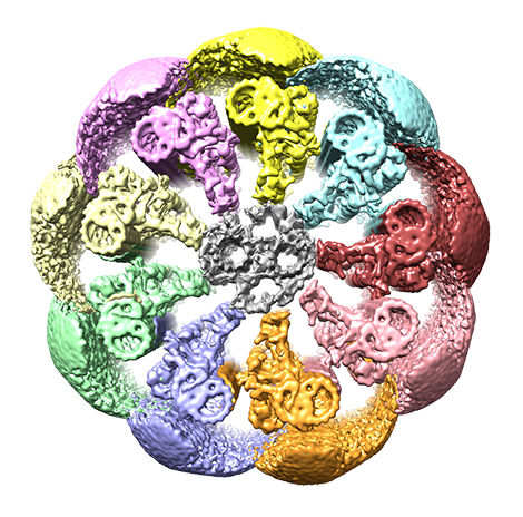
Janelia’s powerful electron microscopes captured images of the protein structures composing the tail of mammalian sperm, revealing how their asymmetric arrangement could lead to the sperm’s 3D movement.
Over hundreds of thousands of years, human sperm have evolved a unique swimming motion to propel them through their watery environment to their ultimate destination: the human egg.

Unlike the sperm of other organisms, whose tail beats up and down in a regular pattern, the tails of human and other mammalian sperm beat irregularly and in 3D, a trait likely benefiting fertilization.
Now, scientists have uncovered the molecules generating this distinctive stroke.
Researchers used the state-of-art electron microscopes at HHMI’s Janelia Research Campus to capture structures of the molecules making up the mammalian sperm’s tail. They discovered mammalian sperm-specific proteins known to control motion arranged asymmetrically in the sperm’s tail. This asymmetric distribution could lead to the uneven, 3D beating observed in mammalian sperm, according to a preprint posted May 16, 2022, on bioRxiv.org from a team led by Janelia’s Executive Director Ron Vale.
The research is the first to describe these proteins and could broaden our understanding of infertility, which affects millions of people every year, says Zhen Chen, a postdoc at the University of California, San Francisco, and lead author of the study. The next step is to determine the molecular identities of these proteins and characterize their function in animal studies. A greater understanding of the molecular mechanism behind sperm motion could potentially reveal new genetic markers for infertility, Chen says.
“Previously, we didn’t even know these proteins were there,” he says. “As they are only found in mammalian sperm, they are likely there to address specific challenges of fertilization in mammals.”
A closer look
The unique motion of mammalian sperm has been observed before, but the molecular structures responsible for the distinctive swimming pattern remained elusive. Only recently have scientists figured out how to image thick sperm samples using electron microscopes, allowing them to peer deep inside the sperm’s tail and see the molecules it comprises.
“We knew that mammalian sperm have different motile behavior compared to other organisms, but we didn’t know the molecules responsible for these differences,” Chen says. “That is why we really wanted to look at the protein structure at the highest possible resolution.”
“We want to know what kind of molecular adaptation had to happen in mammalian sperm to produce the varieties of swimming strokes observed for various mammalian sperm,” he says.
The tail of mammalian sperm is too thick to image on an electron microscope, so the researchers used a cryogenic focused ion beam-scanning electron microscope (cryo-FIB-SEM) to mill away the thick sperm samples to generate slices thin enough for electrons to penetrate. They then used the cryo-electron microscope (cryo-EM) to image the samples using cryo-electron tomography methods (cryo-ET). This combined technique allowed the researchers to capture high-resolution 3D images without altering the proteins.
The team’s experiments started right before COVID-19 affected Janelia’s campus and much of the world. Luckily, Chen was able to stay on campus long enough to work with Momoko Shiozaki and Shumei Zhao, research scientists at Janelia’s cryo-EM and transgenics facilities, to develop a method for preparing and thinning down the samples. Zhiheng Yu, the leader of the cryo-EM facility at Janelia, established methods for remotely collecting data that enabled Chen to work with Janelia scientists virtually to continue the experiments a few months later. Janelia’s cryo-EM facility works to stay at the cutting-edge of the fast-developing fields of cryo-EM and cryo-ET, and help users take advantage of these techniques by implementing new tools, methods, and protocols, Yu says.
Inside the sperm’s tail, or flagella, there are nine microtubules arranged in a circle – a seemingly symmetric structure when viewed in low-resolution images. The researchers found that in mammalian sperm each microtubule is decorated with a distinct combination of protein complexes responsible for regulating the sperm’s motion.
They propose the asymmetric distribution of these protein complexes could make each microtubule move at a different speed and generate a different force. These differences in force could contribute to the unique 3D swimming strokes of mammalian sperm. These movements are likely important for sperm swimming around 3D objects and traveling along the female reproductive tract.
The researchers also found that these proteins are distributed differently in mouse and human sperm.
“These different patterns probably are related to the different way human and mouse sperm are beating,” Chen says. “This reflects the different challenges of human and mouse sperm because, after all, they are swimming in different kinds of environments.”
###
Citation
Zhen Chen, Garrett A. Greenan, Momoko Shiozaki, Yanxin Liu, Will M. Skinner, Xiaowei Zhao, Shumei Zhao, Rui Yan, Caiying Guo, Zhiheng Yu, Polina V. Lishko, David A. Agard, Ronald D. Vale. “In situ cryo-electron tomography reveals the asymmetric architecture of mammalian sperm axonemes.” bioRxiv. Posted May 16, 2022. DOI: 10.1101/2022.05.15.492011
Media Contacts
Nanci Bompey


