
Filter
Associated Lab
- Branson Lab (1) Apply Branson Lab filter
- Dudman Lab (58) Apply Dudman Lab filter
- Harris Lab (1) Apply Harris Lab filter
- Hermundstad Lab (3) Apply Hermundstad Lab filter
- Ji Lab (1) Apply Ji Lab filter
- Karpova Lab (3) Apply Karpova Lab filter
- Lavis Lab (2) Apply Lavis Lab filter
- Lee (Albert) Lab (1) Apply Lee (Albert) Lab filter
- Looger Lab (1) Apply Looger Lab filter
- Pachitariu Lab (2) Apply Pachitariu Lab filter
- Spruston Lab (2) Apply Spruston Lab filter
- Sternson Lab (1) Apply Sternson Lab filter
- Svoboda Lab (3) Apply Svoboda Lab filter
- Tervo Lab (2) Apply Tervo Lab filter
- Tillberg Lab (1) Apply Tillberg Lab filter
Associated Project Team
Publication Date
- 2026 (5) Apply 2026 filter
- 2025 (6) Apply 2025 filter
- 2024 (2) Apply 2024 filter
- 2023 (2) Apply 2023 filter
- 2022 (3) Apply 2022 filter
- 2021 (2) Apply 2021 filter
- 2020 (2) Apply 2020 filter
- 2019 (4) Apply 2019 filter
- 2018 (3) Apply 2018 filter
- 2017 (4) Apply 2017 filter
- 2016 (3) Apply 2016 filter
- 2015 (3) Apply 2015 filter
- 2014 (4) Apply 2014 filter
- 2013 (2) Apply 2013 filter
- 2011 (1) Apply 2011 filter
- 2009 (1) Apply 2009 filter
- 2007 (3) Apply 2007 filter
- 2006 (1) Apply 2006 filter
- 2005 (2) Apply 2005 filter
- 2004 (2) Apply 2004 filter
- 2003 (3) Apply 2003 filter
Type of Publication
58 Publications
Showing 51-58 of 58 resultsThere is strong evidence that synaptic plasticity is a critical cellular mechanism underlying learning and memory. Although the forms of synaptic plasticity used by different circuits and cell types vary, a widespread presumption is that the male and female brain has evolved to use the same form of plasticity within the same circuits during performance on the same task. Here, we used complimentary approaches to determine how activity in the mouse frontal cortex supports the extinction of associative memories in a context-dependent manner. While in vivo recordings show that both male and female mice have similar cue-relevant activity patterns and ensemble dynamics in excitatory neurons from the infralimbic cortex (IL) during learning, activity in amygdala-projecting IL neurons was indispensable for extinction memories only in male mice. Likewise, male but not female mice showed evidence for the recruitment of IL by structural remodeling and clustering of dendritic spines on these neurons, and extinction memory impairments were evident only in male mice after projection-specific IL deletion of the glutamate receptor subunit GRIN2B. This work provides strong evidence that synaptic plasticity mechanisms employed during learning and critical for memory retrieval differ between males and females, which undercuts the utility of one-size-fits all therapeutic approaches for mental health conditions in which memory is disrupted.Competing Interest StatementThe authors have declared no competing interest.
The transformation of synaptic input into patterns of spike output is a fundamental operation that is determined by the particular complement of ion channels that a neuron expresses. Although it is well established that individual ion channel proteins make stochastic transitions between conducting and non-conducting states, most models of synaptic integration are deterministic, and relatively little is known about the functional consequences of interactions between stochastically gating ion channels. Here, we show that a model of stellate neurons from layer II of the medial entorhinal cortex implemented with either stochastic or deterministically gating ion channels can reproduce the resting membrane properties of stellate neurons, but only the stochastic version of the model can fully account for perithreshold membrane potential fluctuations and clustered patterns of spike output that are recorded from stellate neurons during depolarized states. We demonstrate that the stochastic model implements an example of a general mechanism for patterning of neuronal output through activity-dependent changes in the probability of spike firing. Unlike deterministic mechanisms that generate spike patterns through slow changes in the state of model parameters, this general stochastic mechanism does not require retention of information beyond the duration of a single spike and its associated afterhyperpolarization. Instead, clustered patterns of spikes emerge in the stochastic model of stellate neurons as a result of a transient increase in firing probability driven by activation of HCN channels during recovery from the spike afterhyperpolarization. Using this model, we infer conditions in which stochastic ion channel gating may influence firing patterns in vivo and predict consequences of modifications of HCN channel function for in vivo firing patterns.
Enkephalin modulates striatal function, thereby affecting motor performance and addictive behaviors. The proenkephalin gene is also used as a model to study cyclic AMP-mediated gene expression in striatal neurons. The second messenger pathway leading to proenkephalin expression demonstrates how cyclic AMP pathways are synchronized with depolarization. We show that cyclic AMP-mediated regulation of the proenkephalin gene is dependent on the activity of L-type Ca2+ channels. Inhibition of L-type Ca2+ channels blocks forskolin-mediated induction of proenkephalin. The Ca2+-activated kinase, Ca2+/calmodulin kinase, as well as the cyclic AMP-activated kinase, protein kinase A (PKA), are both necessary for the induction of the proenkephalin promoter. Similarly, both kinases are needed for the L-type Ca2+ channel-mediated induction of proenkephalin. This synchronization of second messenger pathways provides a coincidence mechanism that gates proenkephalin synthesis in striatal neurons, ensuring that levels are increased only in the presence of activated PKA and depolarization.
The basal ganglia plays a significant role in transforming activity in the cerebral cortex into directed behavior, involving motor learning, habit formation and the selection of actions based on desirable outcomes, and the organization of the basal ganglia is intimately linked to that of the cerebral cortex. In this chapter, we focus primarily on the neocortical part of the basal ganglia. A general canonical organizational plan of the neocortical-related basal ganglia is described. An understanding of the canonical organization of the neostriatal part of the basal ganglia, provides a framework for determining the general organizational principles of the parts of the basal ganglia connected with allocortical areas and the amygdala, and this is discussed. While it has been proposed that the basal ganglia provide interactions between disparate functional circuits, another approach might be that there are parallel functional circuits, in which distinct functions are for the most part maintained, or segregated, one from the other. This chapter, however, is biased toward the view that there is maintenance of functional parallel circuits in the organization of the basal ganglia, but that the circuit contains neuroanatomical features that provide for considerable interaction between adjacent circuits.
Vertebrates are remarkable for their ability to select and execute goal-directed actions: motor skills critical for thriving in complex, competitive environments. A key aspect of a motor skill is the ability to execute its component movements over a range of speeds, amplitudes and frequencies (vigor). Recent work has indicated that a subcortical circuit, the basal ganglia, is a critical determinant of movement vigor in rodents and primates. We propose that the basal ganglia evolved from a circuit that in lower vertebrates and some mammals is sufficient to directly command simple or stereotyped movements to one that indirectly controls the vigor of goal-directed movements. The implications of a dual role of the basal ganglia in the control of vigor and response to reward are also discussed.
In contrast to our increasingly detailed understanding of how synaptic plasticity provides a cellular substrate for learning and memory, it is less clear how a neuron’s voltage-gated ion channels interact with plastic changes in synaptic strength to influence behavior. We find, using generalized and regional knockout mice, that deletion of the HCN1 channel causes profound motor learning and memory deficits in swimming and rotarod tasks. In cerebellar Purkinje cells, which are a key component of the cerebellar circuit for learning of correctly timed movements, HCN1 mediates an inward current that stabilizes the integrative properties of Purkinje cells and ensures that their input-output function is independent of the previous history of their activity. We suggest that this nonsynaptic integrative function of HCN1 is required for accurate decoding of input patterns and thereby enables synaptic plasticity to appropriately influence the performance of motor activity.
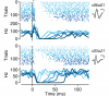
Dysfunction of the basal ganglia produces severe deficits in the timing, initiation, and vigor of movement. These diverse impairments suggest a control system gone awry. In engineered systems, feedback is critical for control. By contrast, models of the basal ganglia highlight feedforward circuitry and ignore intrinsic feedback circuits. In this study, we show that feedback via axon collaterals of substantia nigra projection neurons control the gain of the basal ganglia output. Through a combination of physiology, optogenetics, anatomy, and circuit mapping, we elaborate a general circuit mechanism for gain control in a microcircuit lacking interneurons. Our data suggest that diverse tonic firing rates, weak unitary connections and a spatially diffuse collateral circuit with distinct topography and kinetics from feedforward input is sufficient to implement divisive feedback inhibition. The importance of feedback for engineered systems implies that the intranigral microcircuit, despite its absence from canonical models, could be essential to basal ganglia function. DOI: http://dx.doi.org/10.7554/eLife.02397.001.
Animals adapt their behavior in response to informative sensory cues using multiple brain circuits. The activity of midbrain dopaminergic neurons is thought to convey a critical teaching signal: reward-prediction error. Although reward-prediction error signals are thought to be essential to learning, little is known about the dynamic changes in the activity of midbrain dopaminergic neurons as animals learn about novel sensory cues and appetitive rewards. Here we describe a large dataset of cell-attached recordings of identified dopaminergic neurons as naive mice learned a novel cue-reward association. During learning midbrain dopaminergic neuron activity results from the summation of sensory cue-related and movement initiation-related response components. These components are both a function of reward expectation yet they are dissociable. Learning produces an increasingly precise coordination of action initiation following sensory cues that results in apparent reward-prediction error correlates. Our data thus provide new insights into the circuit mechanisms that underlie a critical computation in a highly conserved learning circuit.