Filter
Associated Lab
- Aguilera Castrejon Lab (19) Apply Aguilera Castrejon Lab filter
- Ahrens Lab (75) Apply Ahrens Lab filter
- Aso Lab (42) Apply Aso Lab filter
- Baker Lab (38) Apply Baker Lab filter
- Betzig Lab (116) Apply Betzig Lab filter
- Beyene Lab (15) Apply Beyene Lab filter
- Bock Lab (17) Apply Bock Lab filter
- Branson Lab (56) Apply Branson Lab filter
- Card Lab (43) Apply Card Lab filter
- Cardona Lab (64) Apply Cardona Lab filter
- Chklovskii Lab (13) Apply Chklovskii Lab filter
- Clapham Lab (16) Apply Clapham Lab filter
- Cui Lab (19) Apply Cui Lab filter
- Darshan Lab (12) Apply Darshan Lab filter
- Dennis Lab (3) Apply Dennis Lab filter
- Dickson Lab (46) Apply Dickson Lab filter
- Druckmann Lab (25) Apply Druckmann Lab filter
- Dudman Lab (58) Apply Dudman Lab filter
- Eddy/Rivas Lab (30) Apply Eddy/Rivas Lab filter
- Egnor Lab (11) Apply Egnor Lab filter
- Espinosa Medina Lab (25) Apply Espinosa Medina Lab filter
- Feliciano Lab (16) Apply Feliciano Lab filter
- Fetter Lab (41) Apply Fetter Lab filter
- FIB-SEM Technology (1) Apply FIB-SEM Technology filter
- Fitzgerald Lab (30) Apply Fitzgerald Lab filter
- Freeman Lab (15) Apply Freeman Lab filter
- Funke Lab (46) Apply Funke Lab filter
- Gonen Lab (91) Apply Gonen Lab filter
- Grigorieff Lab (62) Apply Grigorieff Lab filter
- Harris Lab (65) Apply Harris Lab filter
- Heberlein Lab (94) Apply Heberlein Lab filter
- Hermundstad Lab (32) Apply Hermundstad Lab filter
- Hess Lab (80) Apply Hess Lab filter
- Ilanges Lab (4) Apply Ilanges Lab filter
- Jayaraman Lab (49) Apply Jayaraman Lab filter
- Ji Lab (33) Apply Ji Lab filter
- Johnson Lab (7) Apply Johnson Lab filter
- Kainmueller Lab (19) Apply Kainmueller Lab filter
- Karpova Lab (15) Apply Karpova Lab filter
- Keleman Lab (13) Apply Keleman Lab filter
- Keller Lab (77) Apply Keller Lab filter
- Koay Lab (20) Apply Koay Lab filter
- Lavis Lab (162) Apply Lavis Lab filter
- Lee (Albert) Lab (34) Apply Lee (Albert) Lab filter
- Leonardo Lab (23) Apply Leonardo Lab filter
- Li Lab (32) Apply Li Lab filter
- Lippincott-Schwartz Lab (182) Apply Lippincott-Schwartz Lab filter
- Liu (Yin) Lab (9) Apply Liu (Yin) Lab filter
- Liu (Zhe) Lab (65) Apply Liu (Zhe) Lab filter
- Looger Lab (138) Apply Looger Lab filter
- Magee Lab (49) Apply Magee Lab filter
- Menon Lab (18) Apply Menon Lab filter
- Murphy Lab (13) Apply Murphy Lab filter
- O'Shea Lab (8) Apply O'Shea Lab filter
- Otopalik Lab (13) Apply Otopalik Lab filter
- Pachitariu Lab (56) Apply Pachitariu Lab filter
- Pastalkova Lab (19) Apply Pastalkova Lab filter
- Pavlopoulos Lab (19) Apply Pavlopoulos Lab filter
- Pedram Lab (15) Apply Pedram Lab filter
- Podgorski Lab (16) Apply Podgorski Lab filter
- Reiser Lab (55) Apply Reiser Lab filter
- Riddiford Lab (44) Apply Riddiford Lab filter
- Romani Lab (52) Apply Romani Lab filter
- Rubin Lab (149) Apply Rubin Lab filter
- Saalfeld Lab (66) Apply Saalfeld Lab filter
- Satou Lab (18) Apply Satou Lab filter
- Scheffer Lab (38) Apply Scheffer Lab filter
- Schreiter Lab (72) Apply Schreiter Lab filter
- Schulze Lab (1) Apply Schulze Lab filter
- Sgro Lab (23) Apply Sgro Lab filter
- Shroff Lab (35) Apply Shroff Lab filter
- Simpson Lab (23) Apply Simpson Lab filter
- Singer Lab (80) Apply Singer Lab filter
- Spruston Lab (98) Apply Spruston Lab filter
- Stern Lab (160) Apply Stern Lab filter
- Sternson Lab (54) Apply Sternson Lab filter
- Stringer Lab (44) Apply Stringer Lab filter
- Svoboda Lab (136) Apply Svoboda Lab filter
- Tebo Lab (36) Apply Tebo Lab filter
- Tervo Lab (10) Apply Tervo Lab filter
- Tillberg Lab (22) Apply Tillberg Lab filter
- Tjian Lab (64) Apply Tjian Lab filter
- Truman Lab (88) Apply Truman Lab filter
- Turaga Lab (53) Apply Turaga Lab filter
- Turner Lab (38) Apply Turner Lab filter
- Vale Lab (8) Apply Vale Lab filter
- Voigts Lab (5) Apply Voigts Lab filter
- Wang (Meng) Lab (31) Apply Wang (Meng) Lab filter
- Wang (Shaohe) Lab (25) Apply Wang (Shaohe) Lab filter
- Wong-Campos Lab (1) Apply Wong-Campos Lab filter
- Wu Lab (9) Apply Wu Lab filter
- Zlatic Lab (28) Apply Zlatic Lab filter
- Zuker Lab (25) Apply Zuker Lab filter
Associated Project Team
- CellMap (13) Apply CellMap filter
- COSEM (3) Apply COSEM filter
- FIB-SEM Technology (5) Apply FIB-SEM Technology filter
- Fly Descending Interneuron (12) Apply Fly Descending Interneuron filter
- Fly Functional Connectome (14) Apply Fly Functional Connectome filter
- Fly Olympiad (5) Apply Fly Olympiad filter
- FlyEM (56) Apply FlyEM filter
- FlyLight (50) Apply FlyLight filter
- GENIE (47) Apply GENIE filter
- Integrative Imaging (11) Apply Integrative Imaging filter
- Larval Olympiad (2) Apply Larval Olympiad filter
- MouseLight (18) Apply MouseLight filter
- NeuroSeq (1) Apply NeuroSeq filter
- ThalamoSeq (1) Apply ThalamoSeq filter
- Tool Translation Team (T3) (29) Apply Tool Translation Team (T3) filter
- Transcription Imaging (49) Apply Transcription Imaging filter
Publication Date
- 2026 (118) Apply 2026 filter
- 2025 (223) Apply 2025 filter
- 2024 (209) Apply 2024 filter
- 2023 (158) Apply 2023 filter
- 2022 (192) Apply 2022 filter
- 2021 (194) Apply 2021 filter
- 2020 (196) Apply 2020 filter
- 2019 (202) Apply 2019 filter
- 2018 (232) Apply 2018 filter
- 2017 (217) Apply 2017 filter
- 2016 (209) Apply 2016 filter
- 2015 (252) Apply 2015 filter
- 2014 (236) Apply 2014 filter
- 2013 (194) Apply 2013 filter
- 2012 (190) Apply 2012 filter
- 2011 (190) Apply 2011 filter
- 2010 (161) Apply 2010 filter
- 2009 (158) Apply 2009 filter
- 2008 (140) Apply 2008 filter
- 2007 (106) Apply 2007 filter
- 2006 (92) Apply 2006 filter
- 2005 (67) Apply 2005 filter
- 2004 (57) Apply 2004 filter
- 2003 (58) Apply 2003 filter
- 2002 (39) Apply 2002 filter
- 2001 (28) Apply 2001 filter
- 2000 (29) Apply 2000 filter
- 1999 (14) Apply 1999 filter
- 1998 (18) Apply 1998 filter
- 1997 (16) Apply 1997 filter
- 1996 (10) Apply 1996 filter
- 1995 (18) Apply 1995 filter
- 1994 (12) Apply 1994 filter
- 1993 (10) Apply 1993 filter
- 1992 (6) Apply 1992 filter
- 1991 (11) Apply 1991 filter
- 1990 (11) Apply 1990 filter
- 1989 (6) Apply 1989 filter
- 1988 (1) Apply 1988 filter
- 1987 (7) Apply 1987 filter
- 1986 (4) Apply 1986 filter
- 1985 (5) Apply 1985 filter
- 1984 (2) Apply 1984 filter
- 1983 (2) Apply 1983 filter
- 1982 (3) Apply 1982 filter
- 1981 (3) Apply 1981 filter
- 1980 (1) Apply 1980 filter
- 1979 (1) Apply 1979 filter
- 1976 (2) Apply 1976 filter
- 1973 (1) Apply 1973 filter
- 1970 (1) Apply 1970 filter
- 1967 (1) Apply 1967 filter
Type of Publication
4313 Publications
Showing 2911-2920 of 4313 resultsLive-cell fluorescence light microscopy has emerged as an important tool in the study of cellular biology. The development of fluorescent markers in parallel with super-resolution imaging systems has pushed light microscopy into the realm of molecular visualization at the nanometer scale. Resolutions previously only attained with electron microscopes are now within the grasp of light microscopes. However, until recently, live-cell imaging approaches have eluded super-resolution microscopy, hampering it from reaching its full potential for revealing the dynamic interactions in biology occurring at the single molecule level. Here we examine recent advances in the super-resolution imaging of living cells by reviewing recent breakthroughs in single molecule localization microscopy methods such as PALM and STORM to achieve this important goal.
Different stimulus intensities elicit distinct perceptions, implying that input signals are either conveyed through an overlapping but distinct subpopulation of sensory neurons or channeled into divergent brain circuits according to intensity. In Drosophila, carbon dioxide (CO2) is detected by a single type of olfactory sensory neuron, but information is conveyed to higher brain centers through second-order projection neurons (PNs). Two distinct pathways, PN(v)-1 and PN(v)-2, are necessary and sufficient for avoidance responses to low and high CO2 concentrations, respectively. Whereas low concentrations activate PN(v)-1, high concentrations activate both PN(v)s and GABAergic PN(v)-3, which may inhibit PN(v)-1 pathway-mediated avoidance behavior. Channeling a sensory input into distinct neural pathways allows the perception of an odor to be further modulated by both stimulus intensity and context.
Nervous systems can process information in serial or in parallel, trading off efficiency for flexibility and speed. How these network architectures are implemented across sensorimotor pathways to control behavior is unclear. We investigate this tradeoff directly in Drosophila by comparing neuronal circuits underlying landing and takeoff, behaviors transforming similar visual cues to whole-body motor output. Using a whole-CNS connectome, electrophysiology, and behavioral analysis, we reconstruct the complete feedforward pathway for landing, including visual feature detectors, a dedicated ensemble of descending neurons (DNs), and a core premotor circuit in the nerve cord. Comparison to the takeoff pathway reveals that, despite encoding the same sensory feature and engaging similar muscle groups, neuronal circuits controlling the two behaviors are separated at every sensorimotor level. Extending this analysis to the complete DN population reveals a blueprint for descending motor control: DNs across the behavioral space utilized by the fly are organized as a set of parallel, loosely-overlapping ensembles that form a continuum from command-like control, with individual DNs determining behavioral output, to population coding, with multiple DNs controlling behavior synergistically. Distinct combinations of sensory feature detectors differentially recruit DN ensembles to enable flexible, context-dependent behavioral control.
Ultrasound pulse guided digital phase conjugation has emerged to realize fluorescence imaging inside random scattering media. Its major limitation is the slow imaging speed, as a new wavefront needs to be measured for each voxel. Therefore 3D or even 2D imaging can be time consuming. For practical applications on biological systems, we need to accelerate the imaging process by orders of magnitude. Here we propose and experimentally demonstrate a parallel wavefront measurement scheme towards such a goal. Multiple focused ultrasound pulses of different carrier frequencies can be simultaneously launched inside a scattering medium. Heterodyne interferometry is used to measure all of the wavefronts originating from every sound focus in parallel. We use these wavefronts in sequence to rapidly excite fluorescence at all the voxels defined by the focused ultrasound pulses. In this report, we employed a commercially available sound transducer to generate two sound foci in parallel, doubled the wavefront measurement speed, and reduced the mechanical scanning steps of the sound transducer to half.
A parallel wavefront optimization method is demonstrated experimentally to focus light through random scattering media. The simultaneous modulation of multiple phase elements, each at a unique frequency, enables a parallel determination of the optimal wavefront. Compared to a pixel-by-pixel measurement, the reported parallel method uses the target signal in a highly efficient way. With 441 phase elements, a high-quality focus was formed through a glass diffuser with a peak-to-background ratio of \~{}270. The accuracy and repeatability of the system were tested through experiments.
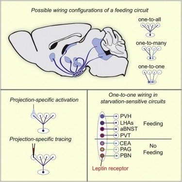
Neural circuits for essential natural behaviors are shaped by selective pressure to coordinate reliable execution of flexible goal-directed actions. However, the structural and functional organization of survival-oriented circuits is poorly understood due to exceptionally complex neuroanatomy. This is exemplified by AGRP neurons, which are a molecularly defined population that is sufficient to rapidly coordinate voracious food seeking and consumption behaviors. Here, we use cell-type-specific techniques for neural circuit manipulation and projection-specific anatomical analysis to examine the organization of this critical homeostatic circuit that regulates feeding. We show that AGRP neuronal circuits use a segregated, parallel, and redundant output configuration. AGRP neuron axon projections that target different brain regions originate from distinct subpopulations, several of which are sufficient to independently evoke feeding. The concerted anatomical and functional analysis of AGRP neuron projection populations reveals a constellation of core forebrain nodes, which are part of an extended circuit that mediates feeding behavior.
We report the experimental studies of a parametric excitation of a second sound (SS) by a first sound (FS) in a superfluid helium in a resonance cavity. The results on several topics in this system are presented: (i) The linear properties of the instability, namely, the threshold, its temperature and geometrical dependencies, and the spectra of SS just above the onset were measured. They were found to be in a good quantitative agreement with the theory. (ii) It was shown that the mechanism of SS amplitude saturation is due to the nonlinear attenuation of SS via three wave interactions between the SS waves. Strong low frequency amplitude fluctuations of SS above the threshold were observed. The spectra of these fluctuations had a universal shape with exponentially decaying tails. Furthermore, the spectral width grew continuously with the FS amplitude. The role of three and four wave interactions are discussed with respect to the nonlinear SS behavior. The first evidence of Gaussian statistics of the wave amplitudes for the parametrically generated wave ensemble was obtained. (iii) The experiments on simultaneous pumping of the FS and independent SS waves revealed new effects. Below the instability threshold, the SS phase conjugation as a result of three-wave interactions between the FS and SS waves was observed. Above the threshold two new effects were found: a giant amplification of the SS wave intensity and strong resonance oscillations of the SS wave amplitude as a function of the FS amplitude. Qualitative explanations of these effects are suggested.
The parasympathetic nervous system helps regulate the functions of many tissues and organs, including the salivary glands and the esophagus. To do so, it needs to reach throughout the body, connecting central systems to peripheral ones. Dyachuk et al. and Espinosa-Medina et al. explored how these connections are established in mice (see the Perspective by Kalcheim and Rohrer). Progenitor cells that travel along with the developing nerves can give rise to both myelinforming Schwann cells and to parasympathetic neurons. That means the interacting nerves do not have to find each other. Instead, the beginnings of the connections are laid down as the nervous system develops. Science, this issue p. 82, p. 87; see also p. 32 Parasympathetic neurons are born from Schwann cell precursors located in the nerves that carry preganglionic fibers. [Also see Perspective by Kalcheim and Rohrer] Neural crest cells migrate extensively and give rise to most of the peripheral nervous system, including sympathetic, parasympathetic, enteric, and dorsal root ganglia. We studied how parasympathetic ganglia form close to visceral organs and what their precursors are. We find that many cranial nerve-associated crest cells coexpress the pan-autonomic determinant Paired-like homeodomain 2b (Phox2b) together with markers of Schwann cell precursors. Some give rise to Schwann cells after down-regulation of PHOX2b. Others form parasympathetic ganglia after being guided to the site of ganglion formation by the nerves that carry preganglionic fibers, a parsimonious way of wiring the pathway. Thus, cranial Schwann cell precursors are the source of parasympathetic neurons during normal development.