Main Menu (Mobile)- Block
Main Menu - Block
SiMView
Quantitative high-speed imaging with Simultaneous Multiview Light-Sheet Microscopy (SiMView)
To obtain the documentation, designs, and parts list, please contact innovation@janelia.hhmi.org.
See additional explanations from the scientists here: https://www.janelia.org/lab/keller-lab/microscopes.
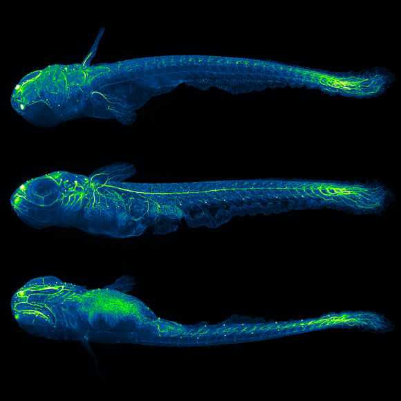
The SiMView microscope acquires multiple views of a specimen simultaneously, using a light-sheet-based design that preserves optimal optical sectioning capability in all views while maximizing physical coverage and imaging speed without compromising sample physiology. This design offers very high imaging speeds of up to several Hz for a volume of 1 cubical millimeter and delivers an excellent spatial resolution of 300 nm laterally and 2 µm axially. At the same time, SiMView microscopy facilitates non-invasive imaging of large-multi-cellular organisms in their entirety for up to several days without the need to sacrifice spatial or temporal resolution. The SiMView microscope is equipped with adaptive imaging capabilities (AutoPilot framework) that allow the microscope to continuously adapt itself to the spatiotemporally variable optical properties of living organisms, thus optimizing spatial resolution and image quality throughout the specimen.
The SiMView microscopy technology framework enables in vivo imaging of entire Drosophila, zebrafish, and mouse embryos at the high speed, resolution, and physical coverage required for comprehensive, automated cell tracking throughout embryogenesis. SiMView microscopy furthermore enables whole-brain functional imaging in larval zebrafish at the single-cell level and whole-animal functional imaging in embryonic and larval Drosophila.
Advantages:
- A powerful tool for live imaging of large biological specimens enabling researchers to understand the development and function of complex biological systems
- exceptionally high imaging speeds, high spatial resolution, and excellent physical coverage of multi-cellular organisms while minimizing photobleaching and phototoxic effects
- vastly superior speed and signal-to-noise ratio compared to confocal and two-photon microscopy
- spatial and temporal artifacts intrinsic to conventional sequential multi-view imaging strategies
Applications:
- Basic life science research, including developmental biology, cell biology, and neurobiology
- Fast volumetric imaging of biological specimens, from single cells to complex multicellular organisms
- Non-invasive live imaging of development with sub-cellular resolution, including entire fruit fly, zebrafish, and mouse embryos
- Non-invasive live imaging of whole-brain activity at the cellular level in the embryonic/larval fruit fly and zebrafish
Publications:
- Single-cell reconstruction of emerging population activity in an entire developing circuit. Wan, Y., Wei, Z., Looger, L.L., Koyama, M., Druckmann, S., and Keller, P.J. Cell, 10.1016/j.cell.2019.08.039 (2019).
- Brain-wide circuit interrogation at the cellular level guided by online analysis of neuronal function. Vladimirov, N., Wang, C., Höckendorf, B., Pujala, A., Tanimoto, M., Mu, Y., Yang, C.-T., Wittenbach, J.D., Freeman, J., Preibisch, S., Koyama, M., Keller, P.J., and Ahrens, M.B. Nature Methods, 15:1117-1125 (2018).
- In toto imaging and reconstruction of post-implantation mouse development at the single-cell level. McDole, K., Guignard, L., Amat, F., Berger, A., Malandain, G., Royer, L.A., Turaga, S.C., Branson, K., and Keller, P.J. Cell, 175:859-876 (2018).
- Adaptive light-sheet microscopy for long-term, high-resolution imaging in living organisms. Royer, L.A., Lemon, W.C., Chhetri, R.K., Wan, Y., Coleman, M., Myers, E.W., and Keller, P.J. Nature Biotechnology, 34:1267-1278 (2016).
- Whole-central nervous system functional imaging in larval Drosophila. Lemon, W.C., Pulver, S.R., Höckendorf, B., McDole, K., Branson, K., Freeman, J., and Keller, P.J. Nature Communications, 6:7924 (2015).
- Fast, accurate reconstruction of cell lineages from large-scale fluorescence microscopy data. Amat, F., Lemon, W.C., Mossing, D., McDole, K., Wan, Y., Branson, K., Myers, E.W., and Keller, P.J. Nature Methods, 11:951-958 (2014).
- Whole-brain functional imaging at cellular resolution using light-sheet microscopy. Ahrens, M.B., Orger, M.B., Robson, D.N., Li, J.M., and Keller, P.J. Nature Methods, 10:413-420 (2013).
- Quantitative high-speed imaging of entire developing embryos with simultaneous multi-view light-sheet microscopy. Tomer, R., Khairy, K., Amat, F., and Keller, P.J. Nature Methods, 9:755-763 (2012).
Patent Status:
Issued U.S. Patent 9,404,869, 10,386,302, and 10,739,266
Opportunity:
Free for Non-Profit Research and available for Commercial License. Please contact Innovation Management at innovation@janelia.hhmi.org.
For inquiries, please reference:
SiMView Microscope, Janelia 2012-013