Main Menu (Mobile)- Block
- Overview
-
Support Teams
- Overview
- Anatomy and Histology
- Cryo-Electron Microscopy
- Electron Microscopy
- Flow Cytometry
- Gene Targeting and Transgenics
- High Performance Computing
- Immortalized Cell Line Culture
- Integrative Imaging
- Invertebrate Shared Resource
- Janelia Experimental Technology
- Mass Spectrometry
- Media Prep
- Molecular Genomics
- Stem Cell & Primary Culture
- Project Pipeline Support
- Project Technical Resources
- Quantitative Genomics
- Scientific Computing
- Viral Tools
- Vivarium
- Open Science
- You + Janelia
- About Us
Main Menu - Block
- Overview
- Anatomy and Histology
- Cryo-Electron Microscopy
- Electron Microscopy
- Flow Cytometry
- Gene Targeting and Transgenics
- High Performance Computing
- Immortalized Cell Line Culture
- Integrative Imaging
- Invertebrate Shared Resource
- Janelia Experimental Technology
- Mass Spectrometry
- Media Prep
- Molecular Genomics
- Stem Cell & Primary Culture
- Project Pipeline Support
- Project Technical Resources
- Quantitative Genomics
- Scientific Computing
- Viral Tools
- Vivarium
Abstract
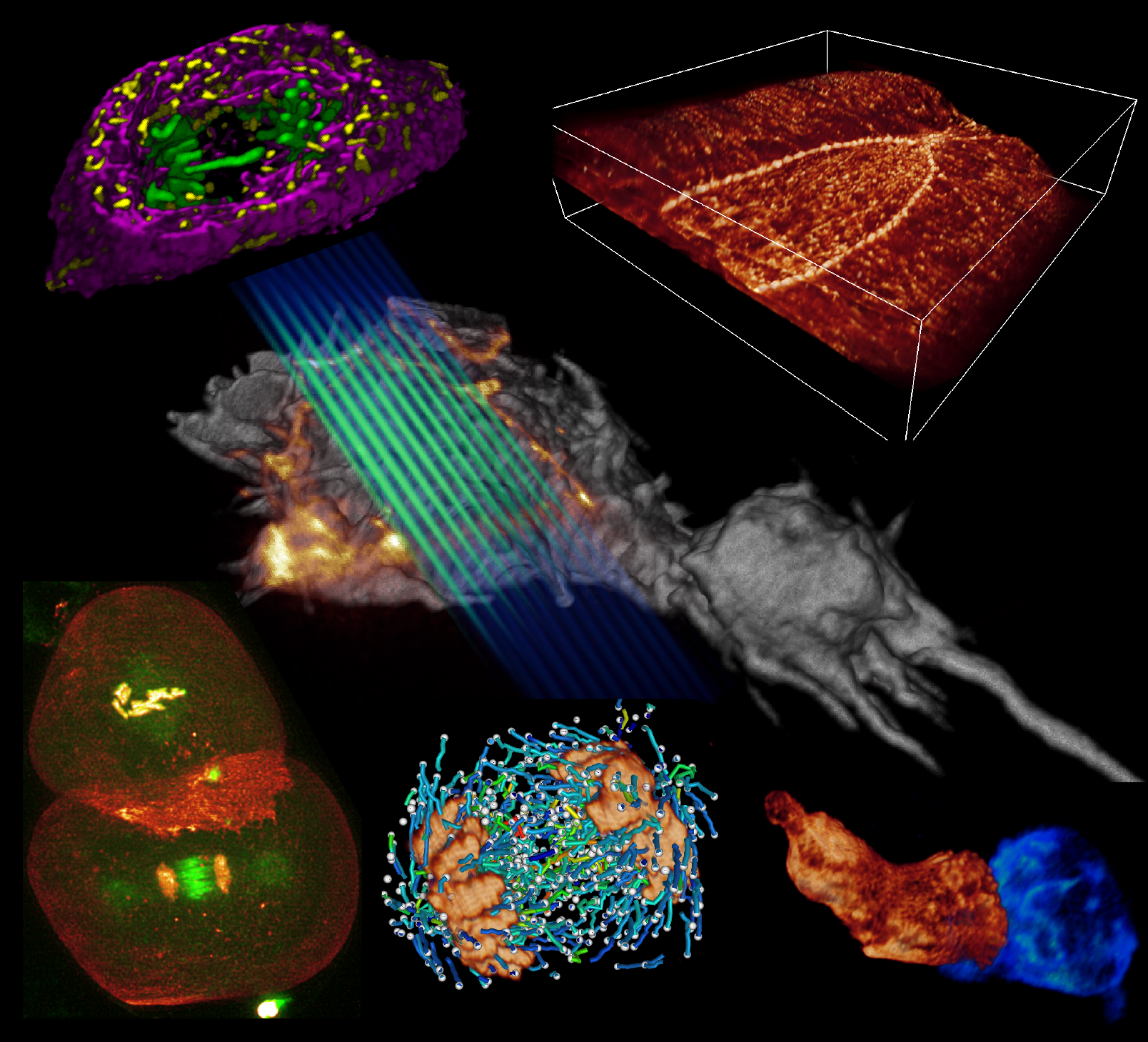
A key challenge when imaging living cells is how to noninvasively extract the most spatiotemporal information possible. Unlike popular wide-field and confocal methods, plane-illumination microscopy limits excitation to the information-rich vicinity of the focal plane, providing effective optical sectioning and high speed while minimizing out-of-focus background and premature photobleaching. Here we used scanned Bessel beams in conjunction with structured illumination and/or two-photon excitation to create thinner light sheets (<0.5 μm) better suited to three-dimensional (3D) subcellular imaging. As demonstrated by imaging the dynamics of mitochondria, filopodia, membrane ruffles, intracellular vesicles and mitotic chromosomes in live cells, the microscope currently offers 3D isotropic resolution down to \~{}0.3 μm, speeds up to nearly 200 image planes per second and the ability to noninvasively acquire hundreds of 3D data volumes from single living cells encompassing tens of thousands of image frames.


