Filter
Associated Lab
- Aguilera Castrejon Lab (4) Apply Aguilera Castrejon Lab filter
- Ahrens Lab (64) Apply Ahrens Lab filter
- Aso Lab (42) Apply Aso Lab filter
- Baker Lab (19) Apply Baker Lab filter
- Betzig Lab (104) Apply Betzig Lab filter
- Beyene Lab (10) Apply Beyene Lab filter
- Bock Lab (14) Apply Bock Lab filter
- Branson Lab (53) Apply Branson Lab filter
- Card Lab (37) Apply Card Lab filter
- Cardona Lab (45) Apply Cardona Lab filter
- Chklovskii Lab (10) Apply Chklovskii Lab filter
- Clapham Lab (15) Apply Clapham Lab filter
- Cui Lab (19) Apply Cui Lab filter
- Darshan Lab (8) Apply Darshan Lab filter
- Dennis Lab (2) Apply Dennis Lab filter
- Dickson Lab (32) Apply Dickson Lab filter
- Druckmann Lab (21) Apply Druckmann Lab filter
- Dudman Lab (46) Apply Dudman Lab filter
- Eddy/Rivas Lab (30) Apply Eddy/Rivas Lab filter
- Egnor Lab (4) Apply Egnor Lab filter
- Espinosa Medina Lab (21) Apply Espinosa Medina Lab filter
- Feliciano Lab (16) Apply Feliciano Lab filter
- Fetter Lab (31) Apply Fetter Lab filter
- FIB-SEM Technology (1) Apply FIB-SEM Technology filter
- Fitzgerald Lab (17) Apply Fitzgerald Lab filter
- Freeman Lab (15) Apply Freeman Lab filter
- Funke Lab (46) Apply Funke Lab filter
- Gonen Lab (59) Apply Gonen Lab filter
- Grigorieff Lab (34) Apply Grigorieff Lab filter
- Harris Lab (55) Apply Harris Lab filter
- Heberlein Lab (13) Apply Heberlein Lab filter
- Hermundstad Lab (28) Apply Hermundstad Lab filter
- Hess Lab (77) Apply Hess Lab filter
- Ilanges Lab (4) Apply Ilanges Lab filter
- Jayaraman Lab (45) Apply Jayaraman Lab filter
- Ji Lab (33) Apply Ji Lab filter
- Johnson Lab (2) Apply Johnson Lab filter
- Karpova Lab (14) Apply Karpova Lab filter
- Keleman Lab (8) Apply Keleman Lab filter
- Keller Lab (62) Apply Keller Lab filter
- Koay Lab (4) Apply Koay Lab filter
- Lavis Lab (150) Apply Lavis Lab filter
- Lee (Albert) Lab (29) Apply Lee (Albert) Lab filter
- Leonardo Lab (19) Apply Leonardo Lab filter
- Li Lab (8) Apply Li Lab filter
- Lippincott-Schwartz Lab (110) Apply Lippincott-Schwartz Lab filter
- Liu (Yin) Lab (4) Apply Liu (Yin) Lab filter
- Liu (Zhe) Lab (60) Apply Liu (Zhe) Lab filter
- Looger Lab (137) Apply Looger Lab filter
- Magee Lab (31) Apply Magee Lab filter
- Menon Lab (12) Apply Menon Lab filter
- Murphy Lab (6) Apply Murphy Lab filter
- O'Shea Lab (7) Apply O'Shea Lab filter
- Otopalik Lab (1) Apply Otopalik Lab filter
- Pachitariu Lab (44) Apply Pachitariu Lab filter
- Pastalkova Lab (6) Apply Pastalkova Lab filter
- Pavlopoulos Lab (7) Apply Pavlopoulos Lab filter
- Pedram Lab (4) Apply Pedram Lab filter
- Podgorski Lab (16) Apply Podgorski Lab filter
- Reiser Lab (49) Apply Reiser Lab filter
- Riddiford Lab (20) Apply Riddiford Lab filter
- Romani Lab (40) Apply Romani Lab filter
- Rubin Lab (111) Apply Rubin Lab filter
- Saalfeld Lab (49) Apply Saalfeld Lab filter
- Satou Lab (3) Apply Satou Lab filter
- Scheffer Lab (38) Apply Scheffer Lab filter
- Schreiter Lab (55) Apply Schreiter Lab filter
- Schulze Lab (1) Apply Schulze Lab filter
- Sgro Lab (3) Apply Sgro Lab filter
- Shroff Lab (35) Apply Shroff Lab filter
- Simpson Lab (18) Apply Simpson Lab filter
- Singer Lab (37) Apply Singer Lab filter
- Spruston Lab (62) Apply Spruston Lab filter
- Stern Lab (77) Apply Stern Lab filter
- Sternson Lab (47) Apply Sternson Lab filter
- Stringer Lab (41) Apply Stringer Lab filter
- Svoboda Lab (132) Apply Svoboda Lab filter
- Tebo Lab (12) Apply Tebo Lab filter
- Tervo Lab (10) Apply Tervo Lab filter
- Tillberg Lab (19) Apply Tillberg Lab filter
- Tjian Lab (17) Apply Tjian Lab filter
- Truman Lab (58) Apply Truman Lab filter
- Turaga Lab (41) Apply Turaga Lab filter
- Turner Lab (27) Apply Turner Lab filter
- Vale Lab (8) Apply Vale Lab filter
- Voigts Lab (5) Apply Voigts Lab filter
- Wang (Meng) Lab (31) Apply Wang (Meng) Lab filter
- Wang (Shaohe) Lab (6) Apply Wang (Shaohe) Lab filter
- Wong-Campos Lab (1) Apply Wong-Campos Lab filter
- Wu Lab (8) Apply Wu Lab filter
- Zlatic Lab (26) Apply Zlatic Lab filter
- Zuker Lab (5) Apply Zuker Lab filter
Associated Project Team
- CellMap (13) Apply CellMap filter
- COSEM (3) Apply COSEM filter
- FIB-SEM Technology (5) Apply FIB-SEM Technology filter
- Fly Descending Interneuron (12) Apply Fly Descending Interneuron filter
- Fly Functional Connectome (14) Apply Fly Functional Connectome filter
- Fly Olympiad (5) Apply Fly Olympiad filter
- FlyEM (56) Apply FlyEM filter
- FlyLight (50) Apply FlyLight filter
- GENIE (47) Apply GENIE filter
- Integrative Imaging (11) Apply Integrative Imaging filter
- Larval Olympiad (2) Apply Larval Olympiad filter
- MouseLight (18) Apply MouseLight filter
- NeuroSeq (1) Apply NeuroSeq filter
- ThalamoSeq (1) Apply ThalamoSeq filter
- Tool Translation Team (T3) (29) Apply Tool Translation Team (T3) filter
- Transcription Imaging (45) Apply Transcription Imaging filter
Associated Support Team
- Project Pipeline Support (5) Apply Project Pipeline Support filter
- Anatomy and Histology (18) Apply Anatomy and Histology filter
- Cryo-Electron Microscopy (49) Apply Cryo-Electron Microscopy filter
- Electron Microscopy (18) Apply Electron Microscopy filter
- Gene Targeting and Transgenics (11) Apply Gene Targeting and Transgenics filter
- High Performance Computing (7) Apply High Performance Computing filter
- Integrative Imaging (25) Apply Integrative Imaging filter
- Invertebrate Shared Resource (40) Apply Invertebrate Shared Resource filter
- Janelia Experimental Technology (40) Apply Janelia Experimental Technology filter
- Management Team (1) Apply Management Team filter
- Mass Spectrometry (1) Apply Mass Spectrometry filter
- Molecular Genomics (15) Apply Molecular Genomics filter
- Project Technical Resources (54) Apply Project Technical Resources filter
- Quantitative Genomics (20) Apply Quantitative Genomics filter
- Scientific Computing (106) Apply Scientific Computing filter
- Stem Cell & Primary Culture (14) Apply Stem Cell & Primary Culture filter
- Viral Tools (14) Apply Viral Tools filter
- Vivarium (7) Apply Vivarium filter
Publication Date
- 2026 (118) Apply 2026 filter
- 2025 (223) Apply 2025 filter
- 2024 (208) Apply 2024 filter
- 2023 (157) Apply 2023 filter
- 2022 (166) Apply 2022 filter
- 2021 (175) Apply 2021 filter
- 2020 (177) Apply 2020 filter
- 2019 (177) Apply 2019 filter
- 2018 (206) Apply 2018 filter
- 2017 (186) Apply 2017 filter
- 2016 (191) Apply 2016 filter
- 2015 (195) Apply 2015 filter
- 2014 (190) Apply 2014 filter
- 2013 (136) Apply 2013 filter
- 2012 (112) Apply 2012 filter
- 2011 (98) Apply 2011 filter
- 2010 (61) Apply 2010 filter
- 2009 (56) Apply 2009 filter
- 2008 (40) Apply 2008 filter
- 2007 (21) Apply 2007 filter
- 2006 (3) Apply 2006 filter
2896 Janelia Publications
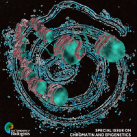
Showing 1651-1660 of 2896 resultsPioneer transcription factors (PTFs) possess the unique capability to access closed chromatin regions and initiate cell fate changes, yet the underlying mechanisms remain elusive. Here, we characterized the single-molecule dynamics of PTFs targeting chromatin in living cells, revealing a notable 'confined target search' mechanism. PTFs such as FOXA1, FOXA2, SOX2, OCT4 and KLF4 sampled chromatin more frequently than non-PTF MYC, alternating between fast free diffusion in the nucleus and slower confined diffusion within mesoscale zones. Super-resolved microscopy showed closed chromatin organized as mesoscale nucleosome-dense domains, confining FOXA2 diffusion locally and enriching its binding. We pinpointed specific histone-interacting disordered regions, distinct from DNA-binding domains, crucial for confined target search kinetics and pioneer activity within closed chromatin. Fusion to other factors enhanced pioneer activity. Kinetic simulations suggested that transient confinement could increase target association rate by shortening search time and binding repeatedly. Our findings illuminate how PTFs recognize and exploit closed chromatin organization to access targets, revealing a pivotal aspect of gene regulation.
Optical nanoscopy of intact biological specimens has been transformed by recent advancements in hydrogel-based tissue clearing and expansion, enabling the imaging of cellular and subcellular structures with molecular contrast. However, existing high-resolution fluorescence microscopes are physically limited by objective-to-specimen distance, which prevents the study of whole-mount specimens without physical sectioning. To address this challenge, we developed a photochemical strategy for spatially precise sectioning of specimens. By combining serial photochemical sectioning with lattice light-sheet imaging and petabyte-scale computation, we imaged and reconstructed axons and myelin sheaths across entire mouse olfactory bulbs at nanoscale resolution. An olfactory bulb–wide analysis of myelinated and unmyelinated axons revealed distinctive patterns of axon degeneration and de-/dysmyelination in the neurodegenerative brain, highlighting the potential for peta- to exabyte-scale super-resolution studies using this approach. High-resolution microscopes have a short working distance, making it difficult to see deep within large biological samples such as an intact brain. Slicing the tissue with a blade can reach deeper, but this often distorts or destroys the fine structures that scientists want to study. By embedding a sample in a light-sensitive hydrogel, Wang et al. demonstrated a gentler approach using a precise ray or sheet of light to dissolve or cut away tissue layer by layer. After each layer is removed, the newly exposed surface is imaged, allowing for a complete, high-resolution, three-dimensional reconstruction without damaging physical contact. bioRxiv preprint: https://www.biorxiv.org/content/10.1101/2024.08.01.605857v1
Seconds-scale network states, affecting many neurons within a network, modulate neural activity by complementing fast integration of neuron-specific inputs that arrive in the milliseconds before spiking. Non-rhythmic subthreshold dynamics at intermediate timescales, however, are less well-characterized. We found, using automated whole cell patch clamping in vivo, that spikes recorded in CA1 and barrel cortex in awake mice are often preceded not only by monotonic voltage rises lasting milliseconds, but also by more gradual (lasting 10s-100s of ms) depolarizations. The latter exert a gating function on spiking, in a fashion that depends on the gradual rise duration: the probability of spiking was higher for longer gradual rises, even controlling for the amplitude of the gradual rises. Barrel cortex double-autopatch recordings show that gradual rises are shared across some but not all neurons. The gradual rises may represent a new kind of state, intermediate both in timescale and in proportion of neurons participating, which gates a neuron's ability to respond to subsequent inputs.
Animals can learn general task structures and use them to solve new problems with novel sensory specifics. This capacity of ‘learning to learn’, or meta-learning, is difficult to achieve in artificial systems, and the mechanisms by which it is achieved in animals are unknown. As a step toward enabling mechanistic studies, we developed a behavioral paradigm that demonstrates meta-learning in head-fixed mice. We trained mice to perform a two-alternative forced-choice task in virtual reality (VR), and successively changed the visual cues that signaled reward location. Mice showed increased learning speed in both cue generalization and serial reversal tasks. During reversal learning, behavior exhibited sharp transitions, with the transition occurring earlier in each successive reversal. Analysis of motor patterns revealed that animals utilized similar motor programs to execute the same actions in response to different cues but modified the motor programs during reversal learning. Our study demonstrates that mice can perform meta-learning tasks in VR, thus opening up opportunities for future mechanistic studies.
Mitochondria-derived reactive oxygen species (mROS) are required for the survival, proliferation, and metastasis of cancer cells. The mechanism by which mitochondrial metabolism regulates mROS levels to support cancer cells is not fully understood. To address this, we conducted a metabolism-focused CRISPR-Cas9 genetic screen and uncovered that loss of genes encoding subunits of mitochondrial complex I was deleterious in the presence of the mitochondria-targeted antioxidant mito-vitamin E (MVE). Genetic or pharmacologic inhibition of mitochondrial complex I in combination with the mitochondria-targeted antioxidants, MVE or MitoTEMPO, induced a robust integrated stress response (ISR) and markedly diminished cell survival and proliferation in vitro. This was not observed following inhibition of mitochondrial complex III. Administration of MitoTEMPO in combination with the mitochondrial complex I inhibitor phenformin decreased the leukemic burden in a mouse model of T cell acute lymphoblastic leukemia. Thus, mitochondrial complex I is a dominant metabolic determinant of mROS-dependent cellular fitness.
The thirteen nuclear cleavages that give rise to the Drosophila blastoderm are some of the fastest known cell cycles. Surprisingly, the fertilized egg is provided with at most one-third of the dNTPs needed to complete the thirteen rounds of DNA replication. The rest must be synthesized by the embryo, concurrent with cleavage divisions. What is the reason for the limited supply of DNA building blocks? We propose that frugal control of dNTP synthesis contributes to the well-characterized deceleration of the cleavage cycles and is needed for robust accumulation of zygotic gene products. In support of this model, we demonstrate that when the levels of dNTPs are abnormally high, nuclear cleavages fail to sufficiently decelerate, the levels of zygotic transcription are dramatically reduced, and the embryo catastrophically fails early in gastrulation. Our work reveals a direct connection between metabolism, the cell cycle, and zygotic transcription.
A specialist neuron uses an intriguing process to help control the body's response to hunger. A lipid pathway involving the breakdown of cellular components regulates the expression of a neuropeptide that affects feeding and body weight.
Insects like Drosophila produce a second brain adapted to the form and behavior of a larva. Neurons for both larval and adult brains are produced by the same stem cells (neuroblasts) but the larva possesses only the earliest born neurons produced from each. To understand how a functional larval brain is made from this reduced set of neurons, we examined the origins and metamorphic fates of the neurons of the larval and adult mushroom body circuits. The adult mushroom body core is built sequentially of γ Kenyon cells, that form a medial lobe, followed by α’β’, and αβ Kenyon cells that form additional medial lobes and two vertical lobes. Extrinsic input (MBINs) and output (MBONs) neurons divide this core into computational compartments. The larval mushroom body contains only γ neurons. Its medial lobe compartments are roughly homologous to those of the adult and same MBONs are used for both. The larval vertical lobe, however, is an analogous “facsimile” that uses a larval-specific branch on the γ neurons to make up for the missing α’β’, and αβ neurons. The extrinsic cells for the facsimile are early-born neurons that trans-differentiate to serve a mushroom body function in the larva and then shift to other brain circuits in the adult. These findings are discussed in the context of the evolution of a larval brain in insects with complete metamorphosis.
The whole-cell patch-clamp method is a gold standard for single-cell analysis of electrical activity, cellular morphology, and gene expression. Prior to our discovery that patch-clamp pipettes could be cleaned and reused, experimental throughput and automation were limited by the need to replace pipettes manually after each experiment. This article presents an optimized protocol for pipette cleaning, which enables it to be performed quickly (< 30 s), resulting in a high yield of whole-cell recording success rate (> 90%) for over 100 reuses of a single pipette. For most patch-clamp experiments (< 30 whole-cell recordings per day), this method enables a single pipette to be used for an entire day of experiments. In addition, we describe easily implementable hardware and software as well as troubleshooting tips to help other labs implement this method in their own experiments. Pipette cleaning enables patch-clamp experiments to be performed with higher throughput, whether manually or in an automated fashion, by eliminating the tedious and skillful task of replacing pipettes. From our experience with numerous electrophysiology laboratories, pipette cleaning can be integrated into existing patch-clamp setups in approximately one day using the hardware and software described in this article. Graphic abstract: Rapid enzymatic cleaning for reuse of patch-clamp pipettes.