Filter
Associated Lab
- Aguilera Castrejon Lab (4) Apply Aguilera Castrejon Lab filter
- Ahrens Lab (62) Apply Ahrens Lab filter
- Aso Lab (42) Apply Aso Lab filter
- Baker Lab (19) Apply Baker Lab filter
- Betzig Lab (104) Apply Betzig Lab filter
- Beyene Lab (10) Apply Beyene Lab filter
- Bock Lab (14) Apply Bock Lab filter
- Branson Lab (52) Apply Branson Lab filter
- Card Lab (37) Apply Card Lab filter
- Cardona Lab (45) Apply Cardona Lab filter
- Chklovskii Lab (10) Apply Chklovskii Lab filter
- Clapham Lab (15) Apply Clapham Lab filter
- Cui Lab (19) Apply Cui Lab filter
- Darshan Lab (8) Apply Darshan Lab filter
- Dennis Lab (2) Apply Dennis Lab filter
- Dickson Lab (32) Apply Dickson Lab filter
- Druckmann Lab (21) Apply Druckmann Lab filter
- Dudman Lab (44) Apply Dudman Lab filter
- Eddy/Rivas Lab (30) Apply Eddy/Rivas Lab filter
- Egnor Lab (4) Apply Egnor Lab filter
- Espinosa Medina Lab (19) Apply Espinosa Medina Lab filter
- Feliciano Lab (12) Apply Feliciano Lab filter
- Fetter Lab (31) Apply Fetter Lab filter
- FIB-SEM Technology (1) Apply FIB-SEM Technology filter
- Fitzgerald Lab (17) Apply Fitzgerald Lab filter
- Freeman Lab (15) Apply Freeman Lab filter
- Funke Lab (46) Apply Funke Lab filter
- Gonen Lab (59) Apply Gonen Lab filter
- Grigorieff Lab (34) Apply Grigorieff Lab filter
- Harris Lab (55) Apply Harris Lab filter
- Heberlein Lab (13) Apply Heberlein Lab filter
- Hermundstad Lab (26) Apply Hermundstad Lab filter
- Hess Lab (77) Apply Hess Lab filter
- Ilanges Lab (4) Apply Ilanges Lab filter
- Jayaraman Lab (44) Apply Jayaraman Lab filter
- Ji Lab (33) Apply Ji Lab filter
- Johnson Lab (1) Apply Johnson Lab filter
- Karpova Lab (13) Apply Karpova Lab filter
- Keleman Lab (8) Apply Keleman Lab filter
- Keller Lab (61) Apply Keller Lab filter
- Koay Lab (3) Apply Koay Lab filter
- Lavis Lab (146) Apply Lavis Lab filter
- Lee (Albert) Lab (29) Apply Lee (Albert) Lab filter
- Leonardo Lab (19) Apply Leonardo Lab filter
- Li Lab (8) Apply Li Lab filter
- Lippincott-Schwartz Lab (108) Apply Lippincott-Schwartz Lab filter
- Liu (Yin) Lab (3) Apply Liu (Yin) Lab filter
- Liu (Zhe) Lab (60) Apply Liu (Zhe) Lab filter
- Looger Lab (137) Apply Looger Lab filter
- Magee Lab (31) Apply Magee Lab filter
- Menon Lab (12) Apply Menon Lab filter
- Murphy Lab (6) Apply Murphy Lab filter
- O'Shea Lab (7) Apply O'Shea Lab filter
- Otopalik Lab (1) Apply Otopalik Lab filter
- Pachitariu Lab (40) Apply Pachitariu Lab filter
- Pastalkova Lab (6) Apply Pastalkova Lab filter
- Pavlopoulos Lab (7) Apply Pavlopoulos Lab filter
- Pedram Lab (4) Apply Pedram Lab filter
- Podgorski Lab (16) Apply Podgorski Lab filter
- Reiser Lab (49) Apply Reiser Lab filter
- Riddiford Lab (20) Apply Riddiford Lab filter
- Romani Lab (39) Apply Romani Lab filter
- Rubin Lab (111) Apply Rubin Lab filter
- Saalfeld Lab (47) Apply Saalfeld Lab filter
- Satou Lab (3) Apply Satou Lab filter
- Scheffer Lab (38) Apply Scheffer Lab filter
- Schreiter Lab (53) Apply Schreiter Lab filter
- Schulze Lab (1) Apply Schulze Lab filter
- Sgro Lab (3) Apply Sgro Lab filter
- Shroff Lab (31) Apply Shroff Lab filter
- Simpson Lab (18) Apply Simpson Lab filter
- Singer Lab (37) Apply Singer Lab filter
- Spruston Lab (62) Apply Spruston Lab filter
- Stern Lab (77) Apply Stern Lab filter
- Sternson Lab (47) Apply Sternson Lab filter
- Stringer Lab (38) Apply Stringer Lab filter
- Svoboda Lab (132) Apply Svoboda Lab filter
- Tebo Lab (11) Apply Tebo Lab filter
- Tervo Lab (9) Apply Tervo Lab filter
- Tillberg Lab (19) Apply Tillberg Lab filter
- Tjian Lab (17) Apply Tjian Lab filter
- Truman Lab (58) Apply Truman Lab filter
- Turaga Lab (41) Apply Turaga Lab filter
- Turner Lab (27) Apply Turner Lab filter
- Vale Lab (8) Apply Vale Lab filter
- Voigts Lab (4) Apply Voigts Lab filter
- Wang (Meng) Lab (29) Apply Wang (Meng) Lab filter
- Wang (Shaohe) Lab (6) Apply Wang (Shaohe) Lab filter
- Wong-Campos Lab (1) Apply Wong-Campos Lab filter
- Wu Lab (8) Apply Wu Lab filter
- Zlatic Lab (26) Apply Zlatic Lab filter
- Zuker Lab (5) Apply Zuker Lab filter
Associated Project Team
- CellMap (13) Apply CellMap filter
- COSEM (3) Apply COSEM filter
- FIB-SEM Technology (5) Apply FIB-SEM Technology filter
- Fly Descending Interneuron (12) Apply Fly Descending Interneuron filter
- Fly Functional Connectome (14) Apply Fly Functional Connectome filter
- Fly Olympiad (5) Apply Fly Olympiad filter
- FlyEM (56) Apply FlyEM filter
- FlyLight (50) Apply FlyLight filter
- GENIE (47) Apply GENIE filter
- Integrative Imaging (9) Apply Integrative Imaging filter
- Larval Olympiad (2) Apply Larval Olympiad filter
- MouseLight (18) Apply MouseLight filter
- NeuroSeq (1) Apply NeuroSeq filter
- ThalamoSeq (1) Apply ThalamoSeq filter
- Tool Translation Team (T3) (29) Apply Tool Translation Team (T3) filter
- Transcription Imaging (45) Apply Transcription Imaging filter
Associated Support Team
- Project Pipeline Support (5) Apply Project Pipeline Support filter
- Anatomy and Histology (18) Apply Anatomy and Histology filter
- Cryo-Electron Microscopy (44) Apply Cryo-Electron Microscopy filter
- Electron Microscopy (18) Apply Electron Microscopy filter
- Gene Targeting and Transgenics (11) Apply Gene Targeting and Transgenics filter
- High Performance Computing (7) Apply High Performance Computing filter
- Integrative Imaging (23) Apply Integrative Imaging filter
- Invertebrate Shared Resource (40) Apply Invertebrate Shared Resource filter
- Janelia Experimental Technology (37) Apply Janelia Experimental Technology filter
- Management Team (1) Apply Management Team filter
- Mass Spectrometry (1) Apply Mass Spectrometry filter
- Molecular Genomics (15) Apply Molecular Genomics filter
- Project Technical Resources (54) Apply Project Technical Resources filter
- Quantitative Genomics (20) Apply Quantitative Genomics filter
- Scientific Computing (103) Apply Scientific Computing filter
- Stem Cell & Primary Culture (14) Apply Stem Cell & Primary Culture filter
- Viral Tools (14) Apply Viral Tools filter
- Vivarium (7) Apply Vivarium filter
Publication Date
- 2026 (70) Apply 2026 filter
- 2025 (222) Apply 2025 filter
- 2024 (209) Apply 2024 filter
- 2023 (157) Apply 2023 filter
- 2022 (166) Apply 2022 filter
- 2021 (175) Apply 2021 filter
- 2020 (177) Apply 2020 filter
- 2019 (177) Apply 2019 filter
- 2018 (206) Apply 2018 filter
- 2017 (186) Apply 2017 filter
- 2016 (191) Apply 2016 filter
- 2015 (195) Apply 2015 filter
- 2014 (190) Apply 2014 filter
- 2013 (136) Apply 2013 filter
- 2012 (112) Apply 2012 filter
- 2011 (98) Apply 2011 filter
- 2010 (61) Apply 2010 filter
- 2009 (56) Apply 2009 filter
- 2008 (40) Apply 2008 filter
- 2007 (21) Apply 2007 filter
- 2006 (3) Apply 2006 filter
2848 Janelia Publications
Showing 1991-2000 of 2848 resultsThe Hippo pathway was originally discovered to control tissue growth in Drosophila and includes the Hippo kinase (Hpo; MST1/2 in mammals), scaffold protein Salvador (Sav; SAV1 in mammals) and the Warts kinase (Wts; LATS1/2 in mammals). The Hpo kinase is activated by binding to Crumbs-Expanded (Crb-Ex) and/or Merlin-Kibra (Mer-Kib) proteins at the apical domain of epithelial cells. Here we show that activation of Hpo also involves the formation of supramolecular complexes with properties of a biomolecular condensate, including concentration dependence and sensitivity to starvation, macromolecular crowding, or 1,6-hexanediol treatment. Overexpressing Ex or Kib induces formation of micron-scale Hpo condensates in the cytoplasm, rather than at the apical membrane. Several Hippo pathway components contain unstructured low-complexity domains and purified Hpo-Sav complexes undergo phase separation in vitro. Formation of Hpo condensates is conserved in human cells. We propose that apical Hpo kinase activation occurs in phase separated "signalosomes" induced by clustering of upstream pathway components.
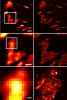
Yes-associated protein (YAP) is a transcriptional co-activator that regulates cell proliferation and survival by binding to a select set of enhancers for target gene activation. How YAP coordinates these transcriptional responses is unknown. Here, we demonstrate that YAP forms liquid-like condensates in the nucleus. Formed within seconds of hyperosmotic stress, YAP condensates compartmentalized the YAP transcription factor TEAD1 and other YAP-related co-activators, including TAZ, and subsequently induced the transcription of YAP-specific proliferation genes. Super-resolution imaging using assay for transposase-accessible chromatin with photoactivated localization microscopy revealed that the YAP nuclear condensates were areas enriched in accessible chromatin domains organized as super-enhancers. Initially devoid of RNA polymerase II, the accessible chromatin domains later acquired RNA polymerase II, transcribing RNA. The removal of the intrinsically-disordered YAP transcription activation domain prevented the formation of YAP condensates and diminished downstream YAP signalling. Thus, dynamic changes in genome organization and gene activation during YAP reprogramming is mediated by liquid-liquid phase separation.
The cricket's auditory system is a highly directional pressure difference receiver whose function is hypothesised to depend on phase relationships between the sound waves propagating through the auditory trachea that connects the left and right hearing organs. We tested this hypothesis by measuring the effect of experimentally constructed phase shifts in acoustic stimuli on phonotactic behavior of Gryllus bimaculatus, the oscillatory response patterns of the tympanic membrane, and the activity of the auditory afferents. The same artificial calling song was played simultaneously at the left and right sides of the cricket, but one sound pattern was shifted in phase by 90 deg (carrier frequencies between 3.6 and 5.4 kHz). All three levels of auditory processing are sensitive to experimentally induced acoustic phase shifts, and the response characteristics are dependent on the carrier frequency of the sound stimulus. At lower frequencies, crickets steered away from the sound leading in phase, while tympanic membrane vibrations and auditory afferent responses were smaller when the ipsilateral sound was leading. In contrast, opposite responses were observed at higher frequencies in all three levels of auditory processing. Minimal responses occurred near the carrier frequency of the cricket's calling song, suggesting a stability at this frequency. Our results indicate that crickets may use directional cues arising from phase shifts in acoustic signals for sound localisation, and that the response properties of pressure difference receivers may be analysed with phase-shifted sound stimuli to further our understanding of how insect auditory systems are adapted for directional processing.
Spectral information plays a crucial role in biological imaging, yet conventional epifluorescence and histological techniques often rely on RGB image acquisition, limiting the resolution of spectrally overlapping components. Here, we present a phasor-based spectral analysis framework adapted for RGB images, enabling unsupervised segmentation and unmixing without the need for hyperspectral systems or sequential acquisition. By applying a discrete Fourier transform to the red, green, and blue intensities at each pixel, we generate a two-dimensional phasor plot where spectral relationships are encoded in modulation and phase. We demonstrate the utility of this approach across three distinct applications: segmentation of lung histology images stained with hematoxylin and eosin to quantify alveolar collapse, analysis of autofluorescence in skin lesions (nevi and melanoma) to highlight pathological spectral signatures, and spectral unmixing in multicolor-labeled U2OS cells to resolve overlapping fluorophores. Our method improves signal separation, reduces noise, and enhances biological interpretability using standard RGB acquisition. These findings establish RGB phasor analysis as a practical and powerful tool for spectral decomposition and segmentation in microscopy, bridging the gap between conventional imaging and advanced spectral analysis.
Mitochondria utilize calcium to increase ATP synthesis. However, excessive matrix calcium activates the mitochondrial permeability transition (mPT), a process that permeabilizes the mitochondrial inner membrane and leads to cell death. While initially characterized 50 y ago, the proteins underlying the process are unclear, although integral membrane proteins were expected to be the porous entities during calcium overload. Here, we designed two assays to study the mPT using high-throughput methodologies. By surveying 19,113 proteins in human cells, we identified four proteins that sensitize the human mPT, but only one that was essential for mPT activation, mitochondrial-localized NRLX1. Surprisingly, NLRX1 is not an integral membrane protein, and our work did not identify any essential integral membrane proteins for the human mPT. The mitochondrial permeability transition (mPT) is an evolutionarily conserved destructive process that permeabilizes the inner mitochondrial membrane in response to calcium overload. The molecular mechanism underlying the mPT is not established. To unambiguously identify essential proteins, we designed two phenotypic assays for mitochondrial calcium overload and applied them to FACS-based CRISPR screening in human cells, ultimately evaluating 19,113 genes. The first screen studied mitochondrial membrane potential (MMP) collapse in response to calcium overload. Top-ranked genes were the essential proteins of the mitochondrial calcium uniporter complex, MCU and EMRE, reflecting that the calcium-induced MMP collapse results from mitochondrial calcium entry and not the mPT. The second screen measured the permeability of the inner mitochondrial membrane. Here, the fluorescent interaction of a membrane impermeant 600 Da dye and a mitochondrial-targeted HaloTag protein was studied under mPT activating conditions; calcium overload and the thiol-reactive molecule phenylarsine oxide. With secondary validation, we identified four protein-encoding genes that delayed or prevented the mPT under knockout: NF2, REST, BPTF, and NRLX1. Knockout of the nonmitochondrial proteins BPTF, NF2, or REST increased mitochondrial calcium retention capacity (CRC). However, calcium release or sensitivity to cyclosporin A (CsA) persisted, indicative of mPT sensitizers. Only knockout of the mitochondrial matrix protein, NLRX1, increased CRC, abolished calcium release, and was CsA-insensitive. This top-ranked hit of the mitochondrial permeability screen meets the definition of an essential mPT activator. Integral membrane proteins, including all previously proposed mPT candidates, were not essential activators.
Proteomic studies have identified thousands of eukaryotic phosphorylation sites (phosphosites), but few are functionally characterized. Nishi et al., in this issue of Structure, characterize phosphosites at protein-protein interfaces and estimate the effect of their phosphorylation on interaction affinity, by combining proteomics data with protein structures.
Photoactivatable pharmacological agents have revolutionized neuroscience, but the palette of available compounds is limited. We describe a general method for caging tertiary amines by using a stable quaternary ammonium linkage that elicits a red shift in the activation wavelength. We prepared a photoactivatable nicotine (PA-Nic), uncageable via one- or two-photon excitation, that is useful to study nicotinic acetylcholine receptors (nAChRs) in different experimental preparations and spatiotemporal scales.
Key to understanding a protein’s biological function is the accurate determination of its spatial distribution inside a cell. Although fluorescent protein markers allow the targeting of specific proteins with molecular precision, much of this information is lost when the resultant fusion proteins are imaged with conventional, diffraction-limited optics. In response, several imaging modalities that are capable of resolution below the diffraction limit (approximately 200 nm) have emerged. Here, both single- and dual-color superresolution imaging of biological structures using photoactivated localization microscopy (PALM) are described. The examples discussed focus on adhesion complexes: dense, protein-filled assemblies that form at the interface between cells and their substrata. A particular emphasis is placed on the instrumentation and photoactivatable fluorescent protein (PA-FP) tags necessary to achieve PALM images at approximately 20 nm resolution in 5 to 30 min in fixed cells.
Commentary: A paper spearheaded by Hari which gives a thorough description of the methods and hardware needed to successfully practice PALM, including cover slip preparation, cell transfection and fixation, drift correction with fiducials, characterization of on/off contrast ratios for different photoactivted fluorescent proteins, identifying PALM-suitable cells, and mechanical and optical components of a PALM system.
A subclass of fluorescent proteins (FPs), large Stokes shift (LSS) FP, are characterized by increased spread between excitation and emission maxima. We report a photoswitchable variant of a red FP with an LSS, PSLSSmKate, which initially exhibits excitation and emission at 445 and 622 nm, but violet irradiation photoswitches PSLSSmKate into a common red form with excitation and emission at 573 and 621 nm. We characterize spectral, photophysical, and biochemical properties of PSLSSmKate in vitro and in mammalian cells and determine its crystal structure in the LSS form. Mass spectrometry, mutagenesis, and spectroscopy of PSLSSmKate allow us to propose molecular mechanisms for the LSS, pH dependence, and light-induced chromophore transformation. We demonstrate the applicability of PSLSSmKate to superresolution photoactivated localization microscopy and protein dynamics in live cells. Given its promising properties, we expect that PSLSSmKate-like phenotype will be further used for photoactivatable imaging and tracking multiple populations of intracellular objects.
Polymorphism is a key feature of amyloid fibril structures but it remains challenging to explain these variations for a particular sample. Here, we report electron cryomicroscopy-based reconstructions from different fibril morphologies formed by a peptide fragment from an amyloidogenic immunoglobulin light chain. The observed fibril morphologies vary in the number and cross-sectional arrangement of a structurally conserved building block. A comparison with the theoretically possible constellations reveals the experimentally observed spectrum of fibril morphologies to be governed by opposing sets of forces that primarily arise from the β-sheet twist, as well as peptide-peptide interactions within the fibril cross-section. Our results provide a framework for rationalizing and predicting the structure and polymorphism of cross-β fibrils, and suggest that a small number of physical parameters control the observed fibril architectures.