Filter
Associated Lab
- Aguilera Castrejon Lab (15) Apply Aguilera Castrejon Lab filter
- Ahrens Lab (56) Apply Ahrens Lab filter
- Aso Lab (39) Apply Aso Lab filter
- Baker Lab (38) Apply Baker Lab filter
- Betzig Lab (110) Apply Betzig Lab filter
- Beyene Lab (10) Apply Beyene Lab filter
- Bock Lab (17) Apply Bock Lab filter
- Branson Lab (48) Apply Branson Lab filter
- Card Lab (40) Apply Card Lab filter
- Cardona Lab (63) Apply Cardona Lab filter
- Chklovskii Lab (13) Apply Chklovskii Lab filter
- Clapham Lab (12) Apply Clapham Lab filter
- Cui Lab (19) Apply Cui Lab filter
- Darshan Lab (12) Apply Darshan Lab filter
- Dennis Lab (1) Apply Dennis Lab filter
- Dickson Lab (46) Apply Dickson Lab filter
- Druckmann Lab (25) Apply Druckmann Lab filter
- Dudman Lab (46) Apply Dudman Lab filter
- Eddy/Rivas Lab (30) Apply Eddy/Rivas Lab filter
- Egnor Lab (11) Apply Egnor Lab filter
- Espinosa Medina Lab (16) Apply Espinosa Medina Lab filter
- Feliciano Lab (6) Apply Feliciano Lab filter
- Fetter Lab (41) Apply Fetter Lab filter
- Fitzgerald Lab (28) Apply Fitzgerald Lab filter
- Freeman Lab (15) Apply Freeman Lab filter
- Funke Lab (34) Apply Funke Lab filter
- Gonen Lab (91) Apply Gonen Lab filter
- Grigorieff Lab (62) Apply Grigorieff Lab filter
- Harris Lab (58) Apply Harris Lab filter
- Heberlein Lab (94) Apply Heberlein Lab filter
- Hermundstad Lab (22) Apply Hermundstad Lab filter
- Hess Lab (71) Apply Hess Lab filter
- Ilanges Lab (1) Apply Ilanges Lab filter
- Jayaraman Lab (44) Apply Jayaraman Lab filter
- Ji Lab (33) Apply Ji Lab filter
- Johnson Lab (6) Apply Johnson Lab filter
- Kainmueller Lab (19) Apply Kainmueller Lab filter
- Karpova Lab (14) Apply Karpova Lab filter
- Keleman Lab (13) Apply Keleman Lab filter
- Keller Lab (75) Apply Keller Lab filter
- Koay Lab (16) Apply Koay Lab filter
- Lavis Lab (136) Apply Lavis Lab filter
- Lee (Albert) Lab (34) Apply Lee (Albert) Lab filter
- Leonardo Lab (23) Apply Leonardo Lab filter
- Li Lab (25) Apply Li Lab filter
- Lippincott-Schwartz Lab (161) Apply Lippincott-Schwartz Lab filter
- Liu (Yin) Lab (5) Apply Liu (Yin) Lab filter
- Liu (Zhe) Lab (58) Apply Liu (Zhe) Lab filter
- Looger Lab (137) Apply Looger Lab filter
- Magee Lab (49) Apply Magee Lab filter
- Menon Lab (18) Apply Menon Lab filter
- Murphy Lab (13) Apply Murphy Lab filter
- O'Shea Lab (4) Apply O'Shea Lab filter
- Otopalik Lab (13) Apply Otopalik Lab filter
- Pachitariu Lab (41) Apply Pachitariu Lab filter
- Pastalkova Lab (18) Apply Pastalkova Lab filter
- Pavlopoulos Lab (19) Apply Pavlopoulos Lab filter
- Pedram Lab (14) Apply Pedram Lab filter
- Podgorski Lab (16) Apply Podgorski Lab filter
- Reiser Lab (49) Apply Reiser Lab filter
- Riddiford Lab (44) Apply Riddiford Lab filter
- Romani Lab (40) Apply Romani Lab filter
- Rubin Lab (139) Apply Rubin Lab filter
- Saalfeld Lab (60) Apply Saalfeld Lab filter
- Satou Lab (16) Apply Satou Lab filter
- Scheffer Lab (36) Apply Scheffer Lab filter
- Schreiter Lab (62) Apply Schreiter Lab filter
- Sgro Lab (20) Apply Sgro Lab filter
- Shroff Lab (23) Apply Shroff Lab filter
- Simpson Lab (23) Apply Simpson Lab filter
- Singer Lab (80) Apply Singer Lab filter
- Spruston Lab (91) Apply Spruston Lab filter
- Stern Lab (152) Apply Stern Lab filter
- Sternson Lab (54) Apply Sternson Lab filter
- Stringer Lab (29) Apply Stringer Lab filter
- Svoboda Lab (135) Apply Svoboda Lab filter
- Tebo Lab (31) Apply Tebo Lab filter
- Tervo Lab (9) Apply Tervo Lab filter
- Tillberg Lab (17) Apply Tillberg Lab filter
- Tjian Lab (64) Apply Tjian Lab filter
- Truman Lab (88) Apply Truman Lab filter
- Turaga Lab (46) Apply Turaga Lab filter
- Turner Lab (35) Apply Turner Lab filter
- Vale Lab (6) Apply Vale Lab filter
- Voigts Lab (2) Apply Voigts Lab filter
- Wang (Meng) Lab (9) Apply Wang (Meng) Lab filter
- Wang (Shaohe) Lab (24) Apply Wang (Shaohe) Lab filter
- Wu Lab (9) Apply Wu Lab filter
- Zlatic Lab (28) Apply Zlatic Lab filter
- Zuker Lab (25) Apply Zuker Lab filter
Associated Project Team
- CellMap (5) Apply CellMap filter
- COSEM (3) Apply COSEM filter
- Fly Descending Interneuron (10) Apply Fly Descending Interneuron filter
- Fly Functional Connectome (14) Apply Fly Functional Connectome filter
- Fly Olympiad (5) Apply Fly Olympiad filter
- FlyEM (51) Apply FlyEM filter
- FlyLight (46) Apply FlyLight filter
- GENIE (40) Apply GENIE filter
- Integrative Imaging (1) Apply Integrative Imaging filter
- Larval Olympiad (2) Apply Larval Olympiad filter
- MouseLight (16) Apply MouseLight filter
- NeuroSeq (1) Apply NeuroSeq filter
- ThalamoSeq (1) Apply ThalamoSeq filter
- Tool Translation Team (T3) (24) Apply Tool Translation Team (T3) filter
- Transcription Imaging (49) Apply Transcription Imaging filter
Publication Date
- 2024 (141) Apply 2024 filter
- 2023 (175) Apply 2023 filter
- 2022 (192) Apply 2022 filter
- 2021 (193) Apply 2021 filter
- 2020 (196) Apply 2020 filter
- 2019 (202) Apply 2019 filter
- 2018 (232) Apply 2018 filter
- 2017 (217) Apply 2017 filter
- 2016 (209) Apply 2016 filter
- 2015 (252) Apply 2015 filter
- 2014 (236) Apply 2014 filter
- 2013 (194) Apply 2013 filter
- 2012 (190) Apply 2012 filter
- 2011 (190) Apply 2011 filter
- 2010 (161) Apply 2010 filter
- 2009 (158) Apply 2009 filter
- 2008 (140) Apply 2008 filter
- 2007 (106) Apply 2007 filter
- 2006 (92) Apply 2006 filter
- 2005 (67) Apply 2005 filter
- 2004 (57) Apply 2004 filter
- 2003 (58) Apply 2003 filter
- 2002 (39) Apply 2002 filter
- 2001 (28) Apply 2001 filter
- 2000 (29) Apply 2000 filter
- 1999 (14) Apply 1999 filter
- 1998 (18) Apply 1998 filter
- 1997 (16) Apply 1997 filter
- 1996 (10) Apply 1996 filter
- 1995 (18) Apply 1995 filter
- 1994 (12) Apply 1994 filter
- 1993 (10) Apply 1993 filter
- 1992 (6) Apply 1992 filter
- 1991 (11) Apply 1991 filter
- 1990 (11) Apply 1990 filter
- 1989 (6) Apply 1989 filter
- 1988 (1) Apply 1988 filter
- 1987 (7) Apply 1987 filter
- 1986 (4) Apply 1986 filter
- 1985 (5) Apply 1985 filter
- 1984 (2) Apply 1984 filter
- 1983 (2) Apply 1983 filter
- 1982 (3) Apply 1982 filter
- 1981 (3) Apply 1981 filter
- 1980 (1) Apply 1980 filter
- 1979 (1) Apply 1979 filter
- 1976 (2) Apply 1976 filter
- 1973 (1) Apply 1973 filter
- 1970 (1) Apply 1970 filter
- 1967 (1) Apply 1967 filter
Type of Publication
3920 Publications
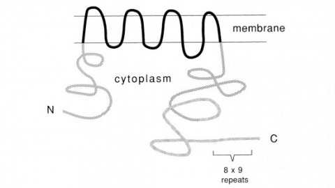
Showing 2251-2260 of 3920 resultsRecent studies suggest that the fly uses the inositol lipid signaling system for visual excitation and that the Drosophila transient receptor potential (trp) mutation disrupts this process subsequent to the production of IP3. In this paper, we show that trp encodes a novel 1275 amino acid protein with eight putative transmembrane segments. Immunolocalization indicates that the trp protein is expressed predominantly in the rhabdomeric membranes of the photoreceptor cells.
Cell recognition requires interactions through molecules located on cell surface. The insect homolog of Down syndrome cell adhesion molecule (Dscam) manifests huge molecular diversity in its extracellular domain. High-affinity Dscam-Dscam interactions only occur between isoforms that carry identical extracellular domains. Homophilic Dscam signaling can, thus, vary in strength depending on the compositions of Dscams present on the opposing cell surfaces. Dscam abundantly exists in the developing nervous system and governs arborization and proper elaboration of neurites. Notably, individual neurons may stochastically and dynamically express a small subset of Dscam isoforms such that any given neurite can be endowed with a unique repertoire of Dscams. This allows individual neurites to recognize their sister branches. Self-recognition leads to self-repulsion, ensuring divergent migration of sister processes. By contrast, weak homophilic Dscam interactions may promote fasciculation of neurites that express analogous, but not identical, Dscams. Differential Dscam binding may provide graded cell recognition that in turn governs complex neuronal morphogenesis.
Glucose transporters (GLUTs) provide a pathway for glucose transport across membranes. Human GLUTs are implicated in devastating diseases such as heart disease, hyper- and hypo-glycemia, type 2 diabetes and caner. The human GLUT1 has been recently crystalized in the inward-facing open conformation. However, there is no other structural information for other conformations. The X-ray structures of E. coli Xylose permease (XylE), a glucose transporter homolog, are available in multiple conformations with and without the substrates D-xylose and D-glucose. XylE has high sequence homology to human GLUT1 and key residues in the sugar-binding pocket are conserved. Here we construct a homology model for human GLUT1 based on the available XylE crystal structure in the partially occluded outward-facing conformation. A long unbiased all atom molecular dynamics simulation starting from the model can capture a new fully opened outward-facing conformation. Our investigation of molecular interactions at the interface between the transmembrane (TM) domains and the intracellular helices (ICH) domain in the outward- and inward-facing conformation supports that the ICH domain likely stabilizes the outward-facing conformation in GLUT1. Furthermore, inducing a conformational transition, our simulations manifest a global asymmetric rocker switch motion and detailed molecular interactions between the substrate and residues through the water-filled selective pore along a pathway from the extracellular to the intracellular side. The results presented here are consistent with previously published biochemical, mutagenesis and functional studies. Together, this study shed light on the structure and functional relationships of GLUT1 in multiple conformational states.
Recently, the fruit fly Drosophila melanogaster has been introduced as a model system to study the molecular bases of a variety of ethanol-induced behaviors. It became immediately apparent that the behavioral changes elicited by acute ethanol exposure are remarkably similar in flies and mammals. Flies show signs of acute intoxication, which range from locomotor stimulation at low doses to complete sedation at higher doses and they develop tolerance upon intermittent ethanol exposure. Genetic screens for mutants with altered responsiveness to ethanol have been carried out and a few of the disrupted genes have been identified. This analysis, while still in its early stages, has already revealed some surprising molecular parallels with mammals. The availability of powerful tools for genetic manipulation in Drosophila, together with the high degree of conservation at the genomic level, make Drosophila a promising model organism to study the mechanism by which ethanol regulates behavior and the mechanisms underlying the organism's adaptation to long-term ethanol exposure.
In Saccharomyces cerevisiae, the repressor Crt1 and the global corepressor Ssn6-Tup1 repress the DNA damage-inducible ribonucleotide reductase (RNR) genes. Initiation of DNA damage signals causes the release of Crt1 and Ssn6-Tup1 from the promoter, coactivator recruitment, and derepression of transcription, indicating that Crt1 plays a crucial role in the switch between gene repression and activation. Here we have mapped the functional domains of Crt1 and identified two independent repression domains and a region required for gene activation. The N terminus of Crt1 is the major repression domain, it directly binds to the Ssn6-Tup1 complex, and its repression activities are dependent upon Ssn6-Tup1 and histone deacetylases (HDACs). In addition, we identified a C-terminal repression domain, which is independent of Ssn6-Tup1 and HDACs and functions at native genes in vivo. Furthermore, we show that TFIID and SWI/SNF bind to a region within the N terminus of Crt1, overlapping with but distinct from the Ssn6-Tup1 binding and repression domain, suggesting that Crt1 may have activator functions. Crt1 mutants were constructed to dissect its activator and repressor functions. All of the mutants were competent for repression of the DNA damage-inducible genes, but a majority were "derepression-defective" mutants. Further characterization of these mutants indicated that they are capable of receiving DNA damage signals and releasing the Ssn6-Tup1 complex from the promoter but are selectively impaired for TFIID and SWI/SNF recruitment. These results imply a two-step activation model of the DNA damage-inducible genes and that Crt1 functions as a signal-dependent dual-transcription activator and repressor that acts in a transient manner.
Rotaviruses, major causes of childhood gastroenteritis, are nonenveloped, icosahedral particles with double-strand RNA genomes. By the use of electron cryomicroscopy and single-particle reconstruction, we have visualized a rotavirus particle comprising the inner capsid coated with the trimeric outer-layer protein, VP7, at a resolution (4 A) comparable with that of X-ray crystallography. We have traced the VP7 polypeptide chain, including parts not seen in its X-ray crystal structure. The 3 well-ordered, 30-residue, N-terminal "arms" of each VP7 trimer grip the underlying trimer of VP6, an inner-capsid protein. Structural differences between free and particle-bound VP7 and between free and VP7-coated inner capsids may regulate mRNA transcription and release. The Ca(2+)-stabilized VP7 intratrimer contact region, which presents important neutralizing epitopes, is unaltered upon capsid binding.
Coordinated motor behaviors depend on feedback communication between peripheral sensory systems and central circuits in the brain and spinal cord. Relay of muscle and tendon-derived sensory information to the CNS is facilitated by functionally and anatomically diverse groups of spinocerebellar tract neurons (SCTNs), but the molecular logic by which SCTN diversity and connectivity is achieved is poorly understood. We used single cell RNA sequencing and genetic manipulations to define the mechanisms governing the molecular profile and organization of SCTN subtypes. We found that SCTNs relaying proprioceptive sensory information from limb and axial muscles are generated through segmentally-restricted actions of specific Hox genes. Loss of Hox function disrupts SCTN subtype-specific transcriptional programs, leading to defects in the connections between proprioceptive sensory neurons, SCTNs, and the cerebellum. These results indicate that Hox-dependent genetic programs play essential roles in the assembly of the neural circuits required for proprioception.
Focal adhesions (FAs) link the extracellular matrix to the actin cytoskeleton to mediate cell adhesion, migration, mechanosensing and signalling. FAs have conserved nanoscale protein organization, suggesting that the position of proteins within FAs regulates their activity and function. Vinculin binds different FA proteins to mediate distinct cellular functions, but how vinculin's interactions are spatiotemporally organized within FAs is unknown. Using interferometric photoactivation localization super-resolution microscopy to assay vinculin nanoscale localization and a FRET biosensor to assay vinculin conformation, we found that upward repositioning within the FA during FA maturation facilitates vinculin activation and mechanical reinforcement of FAs. Inactive vinculin localizes to the lower integrin signalling layer in FAs by binding to phospho-paxillin. Talin binding activates vinculin and targets active vinculin higher in FAs where vinculin can engage retrograde actin flow. Thus, specific protein interactions are spatially segregated within FAs at the nanoscale to regulate vinculin activation and function.
Corticosteroids (CS) act synergistically with thyroid hormone (TH) to accelerate amphibian metamorphosis. Earlier studies showed that CS increase nuclear 3,5,3’-triiodothyronine (T(3)) binding capacity in tadpole tail, and 5’ deiodinase activity in tadpole tissues, increasing the generation of T(3) from thyroxine (T(4)). In the present study we investigated CS synergy with TH by analyzing expression of key genes involved in TH and CS signaling using tadpole tail explant cultures, prometamorphic tadpoles, and frog tissue culture cells (XTC-2 and XLT-15). Treatment of tail explants with T(3) at 100 nM, but not at 10 nM caused tail regression. Corticosterone (CORT) at three doses (100, 500 and 3400 nM) had no effect or increased tail size. T(3) at 10 nM plus CORT caused tails to regress similar to 100 nM T(3). Thyroid hormone receptor beta (TRbeta) mRNA was synergistically upregulated by T(3) plus CORT in tail explants, tail and brain in vivo, and tissue culture cells. The activating 5’ deiodinase type 2 (D2) mRNA was induced by T(3) and CORT in tail explants and tail in vivo. Thyroid hormone increased expression of glucocorticoid (GR) and mineralocorticoid receptor (MR) mRNAs. Our findings support that the synergistic actions of TH and CS in metamorphosis occur at the level of expression of genes for TRbeta and D2, enhancing tissue sensitivity to TH. Concurrently, TH enhances tissue sensitivity to CS by upregulating GR and MR. Environmental stressors can modulate the timing of tadpole metamorphosis in part by CS enhancing the response of tadpole tissues to the actions of TH.

