Filter
Associated Lab
- Dudman Lab (1) Apply Dudman Lab filter
- Harris Lab (7) Apply Harris Lab filter
- Hess Lab (1) Apply Hess Lab filter
- Lee (Albert) Lab (34) Apply Lee (Albert) Lab filter
- Pachitariu Lab (2) Apply Pachitariu Lab filter
- Romani Lab (1) Apply Romani Lab filter
- Spruston Lab (1) Apply Spruston Lab filter
- Sternson Lab (1) Apply Sternson Lab filter
- Svoboda Lab (3) Apply Svoboda Lab filter
Publication Date
- 2023 (5) Apply 2023 filter
- 2022 (1) Apply 2022 filter
- 2021 (1) Apply 2021 filter
- 2020 (5) Apply 2020 filter
- 2019 (1) Apply 2019 filter
- 2018 (2) Apply 2018 filter
- 2017 (6) Apply 2017 filter
- 2016 (1) Apply 2016 filter
- 2014 (4) Apply 2014 filter
- 2012 (2) Apply 2012 filter
- 2011 (1) Apply 2011 filter
- 2010 (1) Apply 2010 filter
- 2009 (1) Apply 2009 filter
- 2006 (1) Apply 2006 filter
- 2004 (1) Apply 2004 filter
- 2002 (1) Apply 2002 filter
Type of Publication
34 Publications
Showing 21-30 of 34 resultsThe hippocampus is critical for recollecting and imagining experiences. This is believed to involve voluntarily drawing from hippocampal memory representations of people, events, and places, including the hippocampus’ map-like representations of familiar environments. However, whether the representations in such “cognitive maps” can be volitionally and selectively accessed is unknown. We developed a brain-machine interface to test if rats could control their hippocampal activity in a flexible, goal-directed, model-based manner. We show that rats can efficiently navigate or direct objects to arbitrary goal locations within a virtual reality arena solely by activating and sustaining appropriate hippocampal representations of remote places. This should provide insight into the mechanisms underlying episodic memory recall, mental simulation/planning, and imagination, and open up possibilities for high-level neural prosthetics utilizing hippocampal representations.
Seconds-scale network states, affecting many neurons within a network, modulate neural activity by complementing fast integration of neuron-specific inputs that arrive in the milliseconds before spiking. Non-rhythmic subthreshold dynamics at intermediate timescales, however, are less well-characterized. We found, using automated whole cell patch clamping in vivo, that spikes recorded in CA1 and barrel cortex in awake mice are often preceded not only by monotonic voltage rises lasting milliseconds, but also by more gradual (lasting 10s-100s of ms) depolarizations. The latter exert a gating function on spiking, in a fashion that depends on the gradual rise duration: the probability of spiking was higher for longer gradual rises, even controlling for the amplitude of the gradual rises. Barrel cortex double-autopatch recordings show that gradual rises are shared across some but not all neurons. The gradual rises may represent a new kind of state, intermediate both in timescale and in proportion of neurons participating, which gates a neuron's ability to respond to subsequent inputs.
Electrophysiology is the most used approach for the collection of functional data in basic and translational neuroscience, but it is typically limited to either intracellular or extracellular recordings. The integration of multiple physiological modalities for the routine acquisition of multimodal data with microelectrodes could be useful for biomedical applications, yet this has been challenging owing to incompatibilities of fabrication methods. Here, we present a suite of glass pipettes with integrated microelectrodes for the simultaneous acquisition of multimodal intracellular and extracellular information in vivo, electrochemistry assessments, and optogenetic perturbations of neural activity. We used the integrated devices to acquire multimodal signals from the CA1 region of the hippocampus in mice and rats, and show that these data can serve as ground-truth validation for the performance of spike-sorting algorithms. The microdevices are applicable for basic and translational neurobiology, and for the development of next-generation brain-machine interfaces.
During many natural behaviors the relevant sensory stimuli and motor outputs are difficult to quantify. Furthermore, the high dimensionality of the space of possible stimuli and movements compounds the problem of experimental control. Head fixation facilitates stimulus control and movement tracking, and can be combined with techniques for recording and manipulating neural activity. However, head-fixed mouse behaviors are typically trained through extensive instrumental conditioning. Here we present a whisker-based, tactile virtual reality system for head-fixed mice running on a spherical treadmill. Head-fixed mice displayed natural movements, including running and rhythmic whisking at 16 Hz. Whisking was centered on a set point that changed in concert with running so that more protracted whisking was correlated with faster running. During turning, whiskers moved in an asymmetric manner, with more retracted whisker positions in the turn direction and protracted whisker movements on the other side. Under some conditions, whisker movements were phase-coupled to strides. We simulated a virtual reality tactile corridor, consisting of two moveable walls controlled in a closed-loop by running speed and direction. Mice used their whiskers to track the walls of the winding corridor without training. Whisker curvature changes, which cause forces in the sensory follicles at the base of the whiskers, were tightly coupled to distance from the walls. Our behavioral system allows for precise control of sensorimotor variables during natural tactile navigation.
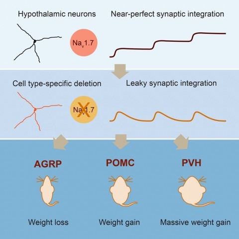
Neurons are well suited for computations on millisecond timescales, but some neuronal circuits set behavioral states over long time periods, such as those involved in energy homeostasis. We found that multiple types of hypothalamic neurons, including those that oppositely regulate body weight, are specialized as near-perfect synaptic integrators that summate inputs over extended timescales. Excitatory postsynaptic potentials (EPSPs) are greatly prolonged, outlasting the neuronal membrane time-constant up to 10-fold. This is due to the voltage-gated sodium channel Nav1.7 (Scn9a), previously associated with pain-sensation but not synaptic integration. Scn9a deletion in AGRP, POMC, or paraventricular hypothalamic neurons reduced EPSP duration, synaptic integration, and altered body weight in mice. In vivo whole-cell recordings in the hypothalamus confirmed near-perfect synaptic integration. These experiments show that integration of synaptic inputs over time by Nav1.7 is critical for body weight regulation and reveal a mechanism for synaptic control of circuits regulating long term homeostatic functions.
Measuring the dynamics of neural processing across time scales requires following the spiking of thousands of individual neurons over milliseconds and months. To address this need, we introduce the Neuropixels 2.0 probe together with newly designed analysis algorithms. The probe has more than 5000 sites and is miniaturized to facilitate chronic implants in small mammals and recording during unrestrained behavior. High-quality recordings over long time scales were reliably obtained in mice and rats in six laboratories. Improved site density and arrangement combined with newly created data processing methods enable automatic post hoc correction for brain movements, allowing recording from the same neurons for more than 2 months. These probes and algorithms enable stable recordings from thousands of sites during free behavior, even in small animals such as mice.
Electrophysiology is one of the major experimental techniques used in neuroscience. The favorable spatial and temporal resolution as well as the increasingly larger site counts of brain recording electrodes contribute to the popularity and importance of electrophysiology in neuroscience. Such electrodes are typically mechanically placed in the brain to perform acute or chronic freely moving animal measurements. The micro positioners currently used for such tasks employ a single translator per independent probe being placed into the targeted brain region, leading to significant size and weight restrictions. To overcome this limitation, we have developed a miniature robotic multi-probe neural microdrive that utilizes novel phase-change-material-filled resistive heater micro-grippers. The microscopic dimensions, gentle gripping action, independent electronic actuation control, and high packing density of the grippers allow for micrometer-precision independent positioning of multiple arbitrarily shaped parallel neural electrodes with only a single piezo actuator in an inchworm motor configuration. This multi-probe-single-actuator design allows for significant size and weight reduction, as well as remote control and potential automation of the microdrive. We demonstrate accurate placement of multiple independent recording electrodes into the CA1 region of the rat hippocampus in vivo in acute and chronic settings. Thus, our robotic neural microdrive technology is applicable towards basic neuroscience and clinical studies, as well as other multi-probe or multi-sensor micro-positioning applications.
The claustrum is one of the most widely connected regions of the forebrain, yet its function has remained obscure, largely due to the experimentally challenging nature of targeting this small, thin, and elongated brain area. However, recent advances in molecular techniques have enabled the anatomy and physiology of the claustrum to be studied with the spatiotemporal and cell type-specific precision required to eventually converge on what this area does. Here we review early anatomical and electrophysiological results from cats and primates, as well as recent work in the rodent, identifying the connectivity, cell types, and physiological circuit mechanisms underlying the communication between the claustrum and the cortex. The emerging picture is one in which the rodent claustrum is closely tied to frontal/limbic regions and plays a role in processes, such as attention, that are associated with these areas. Expected final online publication date for the , Volume 43 is July 8, 2020. Please see http://www.annualreviews.org/page/journal/pubdates for revised estimates.
The claustrum is a brain region that has been investigated for over 200 years, yet its precise function remains unknown. In the final posthumously released article of Francis Crick, written with Christof Koch, the claustrum was suggested to be critically linked to consciousness. Though the claustrum remained relatively obscure throughout the last half century, it has enjoyed a renewed interest in the last 15 years since Crick and Koch's article. During this time, the claustrum, like many other brain regions, has been studied with the myriad of modern systems neuroscience tools that have been made available by the intersection of genetic and viral technologies. This has uncovered new information about its anatomical connectivity and physiological properties and begun to reveal aspects of its function. From these studies, one clear consensus has emerged which supports Crick and Koch's primary interest in the claustrum: the claustrum has widespread extensive connectivity with the entire cerebral cortex, suggesting a prominent role in 'higher order processes'.
Hippocampal activity represents many behaviorally important variables, including context, an animal's location within a given environmental context, time, and reward. Using longitudinal calcium imaging in mice, multiple large virtual environments, and differing reward contingencies, we derived a unified probabilistic model of CA1 representations centered on a single feature-the field propensity. Each cell's propensity governs how many place fields it has per unit space, predicts its reward-related activity, and is preserved across distinct environments and over months. Propensity is broadly distributed-with many low, and some very high, propensity cells-and thus strongly shapes hippocampal representations. This results in a range of spatial codes, from sparse to dense. Propensity varied ∼10-fold between adjacent cells in salt-and-pepper fashion, indicating substantial functional differences within a presumed cell type. Intracellular recordings linked propensity to cell excitability. The stability of each cell's propensity across conditions suggests this fundamental property has anatomical, transcriptional, and/or developmental origins.