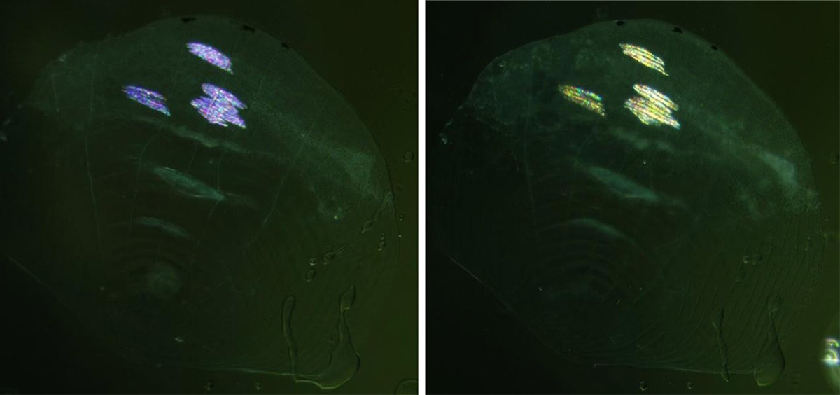
This video shows time-lapse imaging of a zebrafish scale treated with adenosine following treatment with norepinephrine (NE). NE induces the tilting of iridophore crystals and shifts the color from blue to yellow, while adenosine antagonize the NE-induced color change by elevating intracellular cAMP levels. This movie shows that the addition of adenosine causes a rapid reversal of the color change, with the yellow color transitioning back to blue within 10 minutes of treatment. Credit: Gur et al.
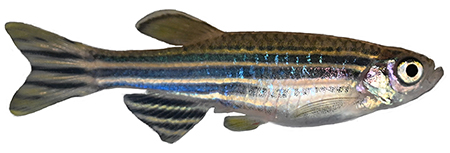
Although zebrafish are much smaller and less famous than their terrestrial namesakes, the tiny fish possess a unique ability: They can rapidly change the color of their characteristic stripes from blue to yellow when they’re distressed.
Like chameleons, zebrafish achieve this color transformation through structural changes. By precisely and simultaneously altering the orientation of light-reflecting crystals on their scales and skin, zebrafish can change the color of their stripes across the entire length of their body in seconds.
 In new research, scientists have pinpointed the intricate cellular machinery behind this color change. Using advanced imaging techniques, they have identified the molecules, structures, and signaling mechanisms inside the cell that work together to change the zebrafish’s stripes from blue to yellow when the fish is under stress.
“Nobody has seen these structures before at this level, and nobody has shown how they respond to changes in light and color,” says Jennifer Lippincott-Schwartz, a senior group leader and head of the 4D Cellular Physiology research area at Janelia, who was the senior author on the new study in collaboration with the lab of John Hammer from the NIH. “There has been a proposal that the crystals are somehow altering their arrangement to change their color, but we are showing precisely how that happens.”
The new findings could help scientists better understand the molecular mechanisms underlying color change in other animals – from chameleons to copepods – that use similar structural color changes to communicate, regulate body temperature, and create camouflage.
“It makes sense that the same components that are needed for this are available and present in other systems, so we think this could be a robust way for organisms to change their color. And we have some preliminary indications that this is actually taking place in other organisms,” says Dvir Gur, a researcher at the Weizmann Institute of Science, who led the new work.
Investigating zebrafish stripes
Gur started examining how zebrafish got their stripes as a postdoctoral researcher in the Lippincott-Schwartz lab. In 2020, Gur, Lippincott-Schwartz, and a team of researchers identified how the ordering of tiny guanine crystals in the zebrafish’s scales generate its blue and yellow stripes.
While studying the fish, the researchers were intrigued by how the zebrafish’s blue stripes would disappear when a fish handler entered the room or when it lost a fight to a dominant male.
In many animals, color changes happen when sacs of pigment disperse and aggregate within the cell. But this wasn’t the case in zebrafish iridophores, where such a movement of the crystals inside these cells would have caused the iridophores to lose their structural-based color. There were hints that the fish could change the orientation of the crystals to reflect light at different angles, creating different colors, but how that happened was not understood.
“This really was the trigger that got us looking at the mechanism that facilitated the color change in these cells,” Gur says. “We knew there had to be another way.”
A close-up look
The team started by getting a closer look at the crystals before and after the color change using high-resolution imaging and synchrotron-based X-ray diffraction.
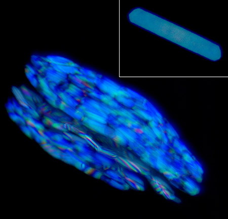
They saw that inside iridophores, the crystals are arranged in stacks of long, plate-like structures. The color change arises from the simultaneous and precise tilting of these crystals. Gur likens the process to the movement of a venetian blind, where the slats tilt together to control how much light comes through. When a zebrafish is stressed, the crystals all tilt at a 20-degree angle, changing the spacing between them and the angle of light hitting them. This alters the optical properties of the crystals in the iridophore, causing the fish’s stripes to change from blue to yellow.

Next, the researchers used live imaging to understand what was driving this process. After artificially inducing a stress response in the fish, the team found that the tilting was enabled by motor proteins called dynein that walk along microtubules inside the cell and connect to the crystals, tugging and tilting them to create the color change. The process is regulated by a molecule called cyclic AMP, a second messenger molecule that is activated when the fish is stressed. Cyclic AMP sends a signal to many cells in the fish at the same time, triggering the tilting and causing all the stripes to change their color simultaneously.
 Beyond providing a mechanism for structural color change, the new findings could help shed light on why some animals form these molecular crystals, which in humans can form kidney stones and gout. They could also inform the design of man-made materials and devices that take advantage of these natural properties.
“For me, it’s really all about curiosity-driven science: Everything that we do is because we want to understand nature better,” says Gur, adding that it is remarkable to see how tiny organisms can achieve something that humans, with their advanced technology, cannot. “But, out of this could come many different things that could eventually also be useful: from using nature as a source to learn principles for biomimicry, to optical devices that are using similar approaches, to next-generation tunable photonic crystals.”
###
Citation:
Dvir Gur, Andrew S. Moore, Rachael Deis, Pang Song, Xufeng Wu, Iddo Pinkas, Claire Deo, Nirmala Iyer, Harald F. Hess, John A. Hammer, and Jennifer Lippincott-Schwartz. “The physical and cellular mechanism of structural color change in zebrafish.” PNAS. Published May 28, 2024. DOI: 10.1073/pnas.2308531121
Media Contacts
Nanci Bompey


