Main Menu (Mobile)- Block
- Overview
-
Support Teams
- Overview
- Anatomy and Histology
- Cryo-Electron Microscopy
- Electron Microscopy
- Flow Cytometry
- Gene Targeting and Transgenics
- High Performance Computing
- Immortalized Cell Line Culture
- Integrative Imaging
- Invertebrate Shared Resource
- Janelia Experimental Technology
- Mass Spectrometry
- Media Prep
- Molecular Genomics
- Primary & iPS Cell Culture
- Project Pipeline Support
- Project Technical Resources
- Quantitative Genomics
- Scientific Computing
- Viral Tools
- Vivarium
- Open Science
- You + Janelia
- About Us
Main Menu - Block
- Overview
- Anatomy and Histology
- Cryo-Electron Microscopy
- Electron Microscopy
- Flow Cytometry
- Gene Targeting and Transgenics
- High Performance Computing
- Immortalized Cell Line Culture
- Integrative Imaging
- Invertebrate Shared Resource
- Janelia Experimental Technology
- Mass Spectrometry
- Media Prep
- Molecular Genomics
- Primary & iPS Cell Culture
- Project Pipeline Support
- Project Technical Resources
- Quantitative Genomics
- Scientific Computing
- Viral Tools
- Vivarium
FLInChR
FLInChR : (Full Length Inverted Channelrhodopsin)
Download our Reagents Catalog here.
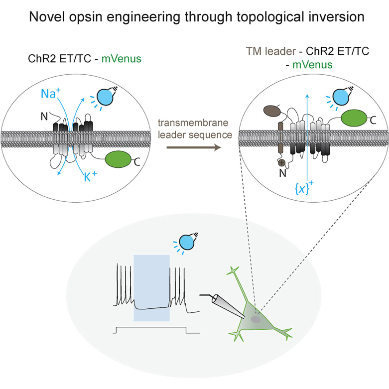
FLInChR is an engineered variant of a light-gated opsin, Channelrhodopsin, that functions as a potent optical inhibitor of neuronal activity. Fusion to the ‘leader’ sequence results in the topological inversion of the opsin and converts it to a light-gated, non-specific cation pump. This ‘flipped’ opsin is efficient as an inhibitor of neuronal activity in both in vitro and in vivo. The ‘topological engineering’ of opsins used to generate FLInChR could serve as a robust platform to screen for variants of membrane-bound proteins with useful properties. Researchers could further use it to expand the optogenetic toolkit.
FLInChR can be expressed in vitro and in vivo in model organisms using DNA constructs (C. elegans) or AAV viral vectors (for rodents). For example, AAV-CAG-FLInChR-mVenus plasmid, distributed by Addgene, can be used to produce viral particles to transduce cells or organisms and express FLInChR in cell types of interest.
Photo stimulation of FLInChR, in vitro and in vivo, is achieved by lasers as described in the publication below (STAR Methods). Further details on the experimental setup are provided in the STAR Methods in the publication.


