Janelia Group Leader Philipp Keller’s laboratory has a knack for finding ways to make the invisible visible. By inventing technology to peer into early vertebrate development, Keller and his team are helping to solve one of biology’s most intriguing problems – how a single cell grows into an animal capable of complex tasks.
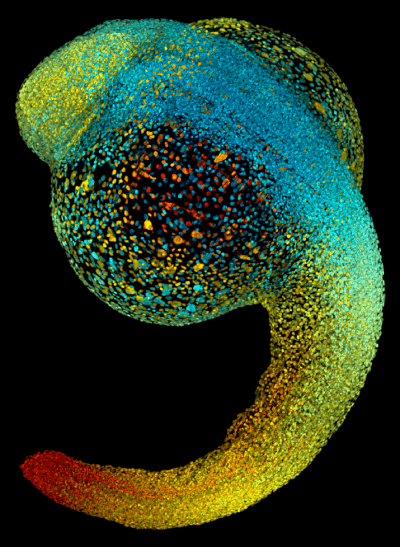
The lab focuses on microscopes that shoot ultrathin laser beams into a living sample, like an embryo, section by section. When paired with powerful software, these light sheet microscopes create movies detailing what happens to each cell throughout development, ultimately revealing the animal’s building plan.
 Light-sheet microscopes let researchers track how individual animal cells, like the ones in this 22-hour old zebrafish embryo, behave during development.Recently, this smart microscope gave researchers the first cellular-level view of a living mouse embryo over 48 hours as its organs took shape and heart cells began to beat. Past work has provided similar details about the early inner workings of zebrafish and fruit flies.
“In the mouse development field, there have been these long-standing questions we haven’t been able to address because we haven’t been able to look at them,” says Kate McDole, a postdoctoral researcher in the Keller Lab who led the work. “The light sheet microscope is the first window that we have into this stage of development. And you can really only do something like that at Janelia, where you not only have the microscope expertise and the tools to build the microscope, but also the collaboration with other labs.”
Accounting for each cell’s behavior during development is essential for scientists striving to regenerate organs or hoping to someday fix developmental problems in the womb.
Such discoveries were recently beyond the limits of imaging technology. The light sheet microscope adapts to rapidly shifting scenes in a way a human technician never could – by making decisions in milliseconds at the cellular level hundreds of times over.
To McDole, the work is a developmental biologist’s equivalent to walking on the moon. “As science has become more niche and detailed, it’s harder and harder to see things that no one’s seen before,” she says.
Keller says Janelia has three qualities that work in concert to help his lab make progress in visualizing vertebrate development. They can tackle difficult work without worrying about the research program’s survival. As they undertake those risky projects, they are also afforded the time to focus on long-term goals and make real breakthroughs. Having a team where physicists, biologists, and computer scientists work together helps them develop meaningful technology and apply it to biological questions.
This all leads to an environment where “you can aim higher and be more ambitious because you have the resources and freedom. In the end, all you need to do is to overcome the scientific challenges,” says Keller.

