Filter
Associated Lab
- Aguilera Castrejon Lab (16) Apply Aguilera Castrejon Lab filter
- Ahrens Lab (64) Apply Ahrens Lab filter
- Aso Lab (40) Apply Aso Lab filter
- Baker Lab (38) Apply Baker Lab filter
- Betzig Lab (112) Apply Betzig Lab filter
- Beyene Lab (13) Apply Beyene Lab filter
- Bock Lab (17) Apply Bock Lab filter
- Branson Lab (52) Apply Branson Lab filter
- Card Lab (40) Apply Card Lab filter
- Cardona Lab (63) Apply Cardona Lab filter
- Chklovskii Lab (13) Apply Chklovskii Lab filter
- Clapham Lab (14) Apply Clapham Lab filter
- Cui Lab (19) Apply Cui Lab filter
- Darshan Lab (12) Apply Darshan Lab filter
- Dennis Lab (1) Apply Dennis Lab filter
- Dickson Lab (46) Apply Dickson Lab filter
- Druckmann Lab (25) Apply Druckmann Lab filter
- Dudman Lab (50) Apply Dudman Lab filter
- Eddy/Rivas Lab (30) Apply Eddy/Rivas Lab filter
- Egnor Lab (11) Apply Egnor Lab filter
- Espinosa Medina Lab (19) Apply Espinosa Medina Lab filter
- Feliciano Lab (7) Apply Feliciano Lab filter
- Fetter Lab (41) Apply Fetter Lab filter
- Fitzgerald Lab (29) Apply Fitzgerald Lab filter
- Freeman Lab (15) Apply Freeman Lab filter
- Funke Lab (38) Apply Funke Lab filter
- Gonen Lab (91) Apply Gonen Lab filter
- Grigorieff Lab (62) Apply Grigorieff Lab filter
- Harris Lab (60) Apply Harris Lab filter
- Heberlein Lab (94) Apply Heberlein Lab filter
- Hermundstad Lab (26) Apply Hermundstad Lab filter
- Hess Lab (76) Apply Hess Lab filter
- Ilanges Lab (2) Apply Ilanges Lab filter
- Jayaraman Lab (46) Apply Jayaraman Lab filter
- Ji Lab (33) Apply Ji Lab filter
- Johnson Lab (6) Apply Johnson Lab filter
- Kainmueller Lab (19) Apply Kainmueller Lab filter
- Karpova Lab (14) Apply Karpova Lab filter
- Keleman Lab (13) Apply Keleman Lab filter
- Keller Lab (76) Apply Keller Lab filter
- Koay Lab (18) Apply Koay Lab filter
- Lavis Lab (148) Apply Lavis Lab filter
- Lee (Albert) Lab (34) Apply Lee (Albert) Lab filter
- Leonardo Lab (23) Apply Leonardo Lab filter
- Li Lab (27) Apply Li Lab filter
- Lippincott-Schwartz Lab (167) Apply Lippincott-Schwartz Lab filter
- Liu (Yin) Lab (6) Apply Liu (Yin) Lab filter
- Liu (Zhe) Lab (61) Apply Liu (Zhe) Lab filter
- Looger Lab (138) Apply Looger Lab filter
- Magee Lab (49) Apply Magee Lab filter
- Menon Lab (18) Apply Menon Lab filter
- Murphy Lab (13) Apply Murphy Lab filter
- O'Shea Lab (6) Apply O'Shea Lab filter
- Otopalik Lab (13) Apply Otopalik Lab filter
- Pachitariu Lab (46) Apply Pachitariu Lab filter
- Pastalkova Lab (18) Apply Pastalkova Lab filter
- Pavlopoulos Lab (19) Apply Pavlopoulos Lab filter
- Pedram Lab (15) Apply Pedram Lab filter
- Podgorski Lab (16) Apply Podgorski Lab filter
- Reiser Lab (51) Apply Reiser Lab filter
- Riddiford Lab (44) Apply Riddiford Lab filter
- Romani Lab (43) Apply Romani Lab filter
- Rubin Lab (143) Apply Rubin Lab filter
- Saalfeld Lab (62) Apply Saalfeld Lab filter
- Satou Lab (16) Apply Satou Lab filter
- Scheffer Lab (36) Apply Scheffer Lab filter
- Schreiter Lab (67) Apply Schreiter Lab filter
- Sgro Lab (20) Apply Sgro Lab filter
- Shroff Lab (29) Apply Shroff Lab filter
- Simpson Lab (23) Apply Simpson Lab filter
- Singer Lab (80) Apply Singer Lab filter
- Spruston Lab (93) Apply Spruston Lab filter
- Stern Lab (156) Apply Stern Lab filter
- Sternson Lab (54) Apply Sternson Lab filter
- Stringer Lab (33) Apply Stringer Lab filter
- Svoboda Lab (135) Apply Svoboda Lab filter
- Tebo Lab (33) Apply Tebo Lab filter
- Tervo Lab (9) Apply Tervo Lab filter
- Tillberg Lab (21) Apply Tillberg Lab filter
- Tjian Lab (64) Apply Tjian Lab filter
- Truman Lab (88) Apply Truman Lab filter
- Turaga Lab (49) Apply Turaga Lab filter
- Turner Lab (37) Apply Turner Lab filter
- Vale Lab (7) Apply Vale Lab filter
- Voigts Lab (3) Apply Voigts Lab filter
- Wang (Meng) Lab (17) Apply Wang (Meng) Lab filter
- Wang (Shaohe) Lab (25) Apply Wang (Shaohe) Lab filter
- Wu Lab (9) Apply Wu Lab filter
- Zlatic Lab (28) Apply Zlatic Lab filter
- Zuker Lab (25) Apply Zuker Lab filter
Associated Project Team
- CellMap (12) Apply CellMap filter
- COSEM (3) Apply COSEM filter
- FIB-SEM Technology (2) Apply FIB-SEM Technology filter
- Fly Descending Interneuron (10) Apply Fly Descending Interneuron filter
- Fly Functional Connectome (14) Apply Fly Functional Connectome filter
- Fly Olympiad (5) Apply Fly Olympiad filter
- FlyEM (53) Apply FlyEM filter
- FlyLight (49) Apply FlyLight filter
- GENIE (45) Apply GENIE filter
- Integrative Imaging (2) Apply Integrative Imaging filter
- Larval Olympiad (2) Apply Larval Olympiad filter
- MouseLight (18) Apply MouseLight filter
- NeuroSeq (1) Apply NeuroSeq filter
- ThalamoSeq (1) Apply ThalamoSeq filter
- Tool Translation Team (T3) (26) Apply Tool Translation Team (T3) filter
- Transcription Imaging (49) Apply Transcription Imaging filter
Publication Date
- 2025 (72) Apply 2025 filter
- 2024 (223) Apply 2024 filter
- 2023 (163) Apply 2023 filter
- 2022 (193) Apply 2022 filter
- 2021 (194) Apply 2021 filter
- 2020 (196) Apply 2020 filter
- 2019 (202) Apply 2019 filter
- 2018 (232) Apply 2018 filter
- 2017 (217) Apply 2017 filter
- 2016 (209) Apply 2016 filter
- 2015 (252) Apply 2015 filter
- 2014 (236) Apply 2014 filter
- 2013 (194) Apply 2013 filter
- 2012 (190) Apply 2012 filter
- 2011 (190) Apply 2011 filter
- 2010 (161) Apply 2010 filter
- 2009 (158) Apply 2009 filter
- 2008 (140) Apply 2008 filter
- 2007 (106) Apply 2007 filter
- 2006 (92) Apply 2006 filter
- 2005 (67) Apply 2005 filter
- 2004 (57) Apply 2004 filter
- 2003 (58) Apply 2003 filter
- 2002 (39) Apply 2002 filter
- 2001 (28) Apply 2001 filter
- 2000 (29) Apply 2000 filter
- 1999 (14) Apply 1999 filter
- 1998 (18) Apply 1998 filter
- 1997 (16) Apply 1997 filter
- 1996 (10) Apply 1996 filter
- 1995 (18) Apply 1995 filter
- 1994 (12) Apply 1994 filter
- 1993 (10) Apply 1993 filter
- 1992 (6) Apply 1992 filter
- 1991 (11) Apply 1991 filter
- 1990 (11) Apply 1990 filter
- 1989 (6) Apply 1989 filter
- 1988 (1) Apply 1988 filter
- 1987 (7) Apply 1987 filter
- 1986 (4) Apply 1986 filter
- 1985 (5) Apply 1985 filter
- 1984 (2) Apply 1984 filter
- 1983 (2) Apply 1983 filter
- 1982 (3) Apply 1982 filter
- 1981 (3) Apply 1981 filter
- 1980 (1) Apply 1980 filter
- 1979 (1) Apply 1979 filter
- 1976 (2) Apply 1976 filter
- 1973 (1) Apply 1973 filter
- 1970 (1) Apply 1970 filter
- 1967 (1) Apply 1967 filter
Type of Publication
4064 Publications
Showing 3961-3970 of 4064 resultsDespite advances in experimental techniques and accumulation of large datasets concerning the composition and properties of the cortex, quantitative modeling of cortical circuits under in-vivo-like conditions remains challenging. Here we report and publicly release a biophysically detailed circuit model of layer 4 in the mouse primary visual cortex, receiving thalamo-cortical visual inputs. The 45,000-neuron model was subjected to a battery of visual stimuli, and results were compared to published work and new in vivo experiments. Simulations reproduced a variety of observations, including effects of optogenetic perturbations. Critical to the agreement between responses in silico and in vivo were the rules of functional synaptic connectivity between neurons. Interestingly, after extreme simplification the model still performed satisfactorily on many measurements, although quantitative agreement with experiments suffered. These results emphasize the importance of functional rules of cortical wiring and enable a next generation of data-driven models of in vivo neural activity and computations.
The ability of insects to learn and navigate to specific locations in the environment has fascinated naturalists for decades. The impressive navigational abilities of ants, bees, wasps and other insects demonstrate that insects are capable of visual place learning, but little is known about the underlying neural circuits that mediate these behaviours. Drosophila melanogaster (common fruit fly) is a powerful model organism for dissecting the neural circuitry underlying complex behaviours, from sensory perception to learning and memory. Drosophila can identify and remember visual features such as size, colour and contour orientation. However, the extent to which they use vision to recall specific locations remains unclear. Here we describe a visual place learning platform and demonstrate that Drosophila are capable of forming and retaining visual place memories to guide selective navigation. By targeted genetic silencing of small subsets of cells in the Drosophila brain, we show that neurons in the ellipsoid body, but not in the mushroom bodies, are necessary for visual place learning. Together, these studies reveal distinct neuroanatomical substrates for spatial versus non-spatial learning, and establish Drosophila as a powerful model for the study of spatial memories.
Visual features detected by the early visual system must be combined into higher order representations to guide behavioral decision. Although key developmental mechanisms that enable the separation of visual feature channels in early visual circuits have been discovered, relatively little is known about the mechanisms that underlie their convergence in later stages of visual processing. Here we explore the development of a functionally well-characterized sensorimotor circuit in Drosophila melanogaster, the convergence of visual projection neurons (VPNs) onto the dendrites of a large descending neuron called the giant fiber (GF). We find two VPNs encoding different visual features that target the same giant fiber dendrite establish their territories on the dendrite, in part, through sequential axon arrival during development prior to synaptogenesis. Physical occupancy is important to maintain territories, as we find the ablation of one VPN results in expanded dendrite territory of the remaining VPN, and that this compensation enables the GF to remain responsive to ethologically relevant visual stimuli. Our data highlight temporal mechanisms for visual feature convergence and promote the GF circuit, and the Drosophila optic glomeruli where VPN to GF connectivity resides, as an ideal developmental model for investigating complex wiring programs and plasticity in visual feature convergence.
Visual projection neurons (VPNs) provide an anatomical connection between early visual processing and higher brain regions. Here we characterize lobula columnar (LC) cells, a class of Drosophila VPNs that project to distinct central brain structures called optic glomeruli. We anatomically describe 22 different LC types and show that, for several types, optogenetic activation in freely moving flies evokes specific behaviors. The activation phenotypes of two LC types closely resemble natural avoidance behaviors triggered by a visual loom. In vivo two-photon calcium imaging reveals that these LC types respond to looming stimuli, while another type does not, but instead responds to the motion of a small object. Activation of LC neurons on only one side of the brain can result in attractive or aversive turning behaviors depending on the cell type. Our results indicate that LC neurons convey information on the presence and location of visual features relevant for specific behaviors.
Many animals rely on vision to detect, locate, and track moving objects. In Drosophila courtship, males primarily use visual cues to orient toward and follow females and to select the ipsilateral wing for courtship song. Here, we show that the LC10 visual projection neurons convey essential visual information during courtship. Males with LC10 neurons silenced are unable to orient toward or maintain proximity to the female and do not predominantly use the ipsilateral wing when singing. LC10 neurons preferentially respond to small moving objects using an antagonistic motion-based center-surround mechanism. Unilateral activation of LC10 neurons recapitulates the orienting and ipsilateral wing extension normally elicited by females, and the potency with which LC10 induces wing extension is enhanced in a state of courtship arousal controlled by male-specific P1 neurons. These data suggest that LC10 is a major pathway relaying visual input to the courtship circuits in the male brain.
Drosophila melanogaster flies cross surmountable gaps in their walkway of widths exceeding their body length with an astounding maneuver but avoid attempts at insurmountable gaps by visual width estimation. Different mutant lines affect specific aspects of this maneuver, indicating a high complexity and modularity of the underlying motor control. Here we report on two mutants, ocelliless(1) and tay bridge(1), that, although making a correct decision to climb, fail dramatically in aiming at the right direction. Both mutants show structural defects in the protocerebral bridge, a central complex neuropil formed like a handlebar spanning the brain hemispheres. The bridge has been implicated in step-length control in walking flies and celestial E-vector orientation in locusts. In rescue experiments using tay bridge(1) flies, the integrity of the bridge was reestablished, concomitantly leading to a significant improvement of their orientation at the gap. Although producing directional scatter, their attempts were clearly aimed at the landing site. However, this partial rescue was lost in these flies at a reduced-visibility landing site. We therefore conclude that the protocerebral bridge is an essential part of a visual targeting network that transmits directional clues to the motor output via a known projection system.
In a wide range of biological studies, it is highly desirable to visualize and analyze three-dimensional (3D) microscopic images. In this primer, we first introduce several major methods for visualizing typical 3D images and related multi-scale, multi-time-point, multi-color data sets. Then, we discuss three key categories of image analysis tasks, namely segmentation, registration, and annotation. We demonstrate how to pipeline these visualization and analysis modules using examples of profiling the single-cell gene-expression of C. elegans and constructing a map of stereotyped neurite tracts in a fruit fly brain.
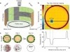
The simultaneous visualization, identification and targeting of neurons during patch clamp-mediated electrophysiological recordings is a basic technique in neuroscience, yet it is often complicated by the inability to visualize the pipette tip, particularly in deep brain tissue. Here we demonstrate a novel approach in which fluorescent quantum dot probes are used to coat pipettes prior to their use. The strong two-photon absorption cross sections of the quantum dots afford robust contrast at significantly deeper penetration depths than current methods allow. We demonstrate the utility of this technique in multiple recording formats both in vitro and in vivo where imaging of the pipettes is achieved at remarkable depths (up to 800 microns). Notably, minimal perturbation of cellular physiology is observed over the hours-long time course of neuronal recordings. We discuss our results within the context of the role that quantum dot nanoprobes may play in understanding neuronal cell physiology.
Neurobiologists investigate the brain of the common fruit fly Drosophila melanogaster to discover neural circuits and link them to complex behaviour. Formulating new hypotheses about connectivity requires potential connectivity information between individual neurons, indicated by overlaps of arborizations of two or more neurons. As the number of higher order overlaps (i.e. overlaps of three or more arborizations) increases exponentially with the number of neurons under investigation, visualization is impeded by clutter and quantification becomes a burden. Existing solutions are restricted to visual or quantitative analysis of pairwise overlaps, as they rely on precomputed overlap data. We present a novel tool that complements existing methods for potential connectivity exploration by providing for the first time the possibility to compute and visualize higher order arborization overlaps on the fly and to interactively explore this information in both its spatial anatomical context and on a quantitative level. Qualitative evaluation by neuroscientists and non-experts demonstrated the utility and usability of the tool.
The transcription and transport of messenger RNA (mRNA) are critical steps in regulating the spatial and temporal components of gene expression, but it has not been possible to observe the dynamics of endogenous mRNA in primary mammalian tissues. We have developed a transgenic mouse in which all β-actin mRNA is fluorescently labeled. We found that β-actin mRNA in primary fibroblasts localizes predominantly by diffusion and trapping as single mRNAs. In cultured neurons and acute brain slices, we found that multiple β-actin mRNAs can assemble together, travel by active transport, and disassemble upon depolarization by potassium chloride. Imaging of brain slices revealed immediate early induction of β-actin transcription after depolarization. Studying endogenous mRNA in live mouse tissues provides insight into its dynamic regulation within the context of the cellular and tissue microenvironment.