Filter
Associated Lab
- Aguilera Castrejon Lab (17) Apply Aguilera Castrejon Lab filter
- Ahrens Lab (69) Apply Ahrens Lab filter
- Aso Lab (42) Apply Aso Lab filter
- Baker Lab (38) Apply Baker Lab filter
- Betzig Lab (115) Apply Betzig Lab filter
- Beyene Lab (14) Apply Beyene Lab filter
- Bock Lab (17) Apply Bock Lab filter
- Branson Lab (54) Apply Branson Lab filter
- Card Lab (43) Apply Card Lab filter
- Cardona Lab (64) Apply Cardona Lab filter
- Chklovskii Lab (13) Apply Chklovskii Lab filter
- Clapham Lab (15) Apply Clapham Lab filter
- Cui Lab (19) Apply Cui Lab filter
- Darshan Lab (12) Apply Darshan Lab filter
- Dennis Lab (2) Apply Dennis Lab filter
- Dickson Lab (46) Apply Dickson Lab filter
- Druckmann Lab (25) Apply Druckmann Lab filter
- Dudman Lab (53) Apply Dudman Lab filter
- Eddy/Rivas Lab (30) Apply Eddy/Rivas Lab filter
- Egnor Lab (11) Apply Egnor Lab filter
- Espinosa Medina Lab (21) Apply Espinosa Medina Lab filter
- Feliciano Lab (10) Apply Feliciano Lab filter
- Fetter Lab (41) Apply Fetter Lab filter
- FIB-SEM Technology (1) Apply FIB-SEM Technology filter
- Fitzgerald Lab (29) Apply Fitzgerald Lab filter
- Freeman Lab (15) Apply Freeman Lab filter
- Funke Lab (42) Apply Funke Lab filter
- Gonen Lab (91) Apply Gonen Lab filter
- Grigorieff Lab (62) Apply Grigorieff Lab filter
- Harris Lab (64) Apply Harris Lab filter
- Heberlein Lab (94) Apply Heberlein Lab filter
- Hermundstad Lab (30) Apply Hermundstad Lab filter
- Hess Lab (79) Apply Hess Lab filter
- Ilanges Lab (3) Apply Ilanges Lab filter
- Jayaraman Lab (48) Apply Jayaraman Lab filter
- Ji Lab (33) Apply Ji Lab filter
- Johnson Lab (6) Apply Johnson Lab filter
- Kainmueller Lab (19) Apply Kainmueller Lab filter
- Karpova Lab (14) Apply Karpova Lab filter
- Keleman Lab (13) Apply Keleman Lab filter
- Keller Lab (76) Apply Keller Lab filter
- Koay Lab (18) Apply Koay Lab filter
- Lavis Lab (154) Apply Lavis Lab filter
- Lee (Albert) Lab (34) Apply Lee (Albert) Lab filter
- Leonardo Lab (23) Apply Leonardo Lab filter
- Li Lab (30) Apply Li Lab filter
- Lippincott-Schwartz Lab (178) Apply Lippincott-Schwartz Lab filter
- Liu (Yin) Lab (7) Apply Liu (Yin) Lab filter
- Liu (Zhe) Lab (64) Apply Liu (Zhe) Lab filter
- Looger Lab (138) Apply Looger Lab filter
- Magee Lab (49) Apply Magee Lab filter
- Menon Lab (18) Apply Menon Lab filter
- Murphy Lab (13) Apply Murphy Lab filter
- O'Shea Lab (7) Apply O'Shea Lab filter
- Otopalik Lab (13) Apply Otopalik Lab filter
- Pachitariu Lab (49) Apply Pachitariu Lab filter
- Pastalkova Lab (18) Apply Pastalkova Lab filter
- Pavlopoulos Lab (19) Apply Pavlopoulos Lab filter
- Pedram Lab (15) Apply Pedram Lab filter
- Podgorski Lab (16) Apply Podgorski Lab filter
- Reiser Lab (54) Apply Reiser Lab filter
- Riddiford Lab (44) Apply Riddiford Lab filter
- Romani Lab (49) Apply Romani Lab filter
- Rubin Lab (148) Apply Rubin Lab filter
- Saalfeld Lab (64) Apply Saalfeld Lab filter
- Satou Lab (16) Apply Satou Lab filter
- Scheffer Lab (38) Apply Scheffer Lab filter
- Schreiter Lab (69) Apply Schreiter Lab filter
- Sgro Lab (21) Apply Sgro Lab filter
- Shroff Lab (31) Apply Shroff Lab filter
- Simpson Lab (23) Apply Simpson Lab filter
- Singer Lab (80) Apply Singer Lab filter
- Spruston Lab (97) Apply Spruston Lab filter
- Stern Lab (158) Apply Stern Lab filter
- Sternson Lab (54) Apply Sternson Lab filter
- Stringer Lab (39) Apply Stringer Lab filter
- Svoboda Lab (135) Apply Svoboda Lab filter
- Tebo Lab (35) Apply Tebo Lab filter
- Tervo Lab (9) Apply Tervo Lab filter
- Tillberg Lab (21) Apply Tillberg Lab filter
- Tjian Lab (64) Apply Tjian Lab filter
- Truman Lab (88) Apply Truman Lab filter
- Turaga Lab (53) Apply Turaga Lab filter
- Turner Lab (39) Apply Turner Lab filter
- Vale Lab (8) Apply Vale Lab filter
- Voigts Lab (4) Apply Voigts Lab filter
- Wang (Meng) Lab (27) Apply Wang (Meng) Lab filter
- Wang (Shaohe) Lab (25) Apply Wang (Shaohe) Lab filter
- Wu Lab (9) Apply Wu Lab filter
- Zlatic Lab (28) Apply Zlatic Lab filter
- Zuker Lab (25) Apply Zuker Lab filter
Associated Project Team
- CellMap (12) Apply CellMap filter
- COSEM (3) Apply COSEM filter
- FIB-SEM Technology (5) Apply FIB-SEM Technology filter
- Fly Descending Interneuron (12) Apply Fly Descending Interneuron filter
- Fly Functional Connectome (14) Apply Fly Functional Connectome filter
- Fly Olympiad (5) Apply Fly Olympiad filter
- FlyEM (56) Apply FlyEM filter
- FlyLight (50) Apply FlyLight filter
- GENIE (47) Apply GENIE filter
- Integrative Imaging (7) Apply Integrative Imaging filter
- Larval Olympiad (2) Apply Larval Olympiad filter
- MouseLight (18) Apply MouseLight filter
- NeuroSeq (1) Apply NeuroSeq filter
- ThalamoSeq (1) Apply ThalamoSeq filter
- Tool Translation Team (T3) (28) Apply Tool Translation Team (T3) filter
- Transcription Imaging (49) Apply Transcription Imaging filter
Publication Date
- 2025 (227) Apply 2025 filter
- 2024 (212) Apply 2024 filter
- 2023 (158) Apply 2023 filter
- 2022 (192) Apply 2022 filter
- 2021 (194) Apply 2021 filter
- 2020 (196) Apply 2020 filter
- 2019 (202) Apply 2019 filter
- 2018 (232) Apply 2018 filter
- 2017 (217) Apply 2017 filter
- 2016 (209) Apply 2016 filter
- 2015 (252) Apply 2015 filter
- 2014 (236) Apply 2014 filter
- 2013 (194) Apply 2013 filter
- 2012 (190) Apply 2012 filter
- 2011 (190) Apply 2011 filter
- 2010 (161) Apply 2010 filter
- 2009 (158) Apply 2009 filter
- 2008 (140) Apply 2008 filter
- 2007 (106) Apply 2007 filter
- 2006 (92) Apply 2006 filter
- 2005 (67) Apply 2005 filter
- 2004 (57) Apply 2004 filter
- 2003 (58) Apply 2003 filter
- 2002 (39) Apply 2002 filter
- 2001 (28) Apply 2001 filter
- 2000 (29) Apply 2000 filter
- 1999 (14) Apply 1999 filter
- 1998 (18) Apply 1998 filter
- 1997 (16) Apply 1997 filter
- 1996 (10) Apply 1996 filter
- 1995 (18) Apply 1995 filter
- 1994 (12) Apply 1994 filter
- 1993 (10) Apply 1993 filter
- 1992 (6) Apply 1992 filter
- 1991 (11) Apply 1991 filter
- 1990 (11) Apply 1990 filter
- 1989 (6) Apply 1989 filter
- 1988 (1) Apply 1988 filter
- 1987 (7) Apply 1987 filter
- 1986 (4) Apply 1986 filter
- 1985 (5) Apply 1985 filter
- 1984 (2) Apply 1984 filter
- 1983 (2) Apply 1983 filter
- 1982 (3) Apply 1982 filter
- 1981 (3) Apply 1981 filter
- 1980 (1) Apply 1980 filter
- 1979 (1) Apply 1979 filter
- 1976 (2) Apply 1976 filter
- 1973 (1) Apply 1973 filter
- 1970 (1) Apply 1970 filter
- 1967 (1) Apply 1967 filter
Type of Publication
4202 Publications
Showing 3101-3110 of 4202 resultsPeer review is an important part of the scientific process, but traditional peer review at journals is coming under increased scrutiny for its inefficiency and lack of transparency. As preprints become more widely used and accepted, they raise the possibility of rethinking the peer-review process. Preprints are enabling new forms of peer review that have the potential to be more thorough, inclusive, and collegial than traditional journal peer review, and to thus fundamentally shift the culture of peer review toward constructive collaboration. In this Consensus View, we make a call to action to stakeholders in the community to accelerate the growing momentum of preprint sharing and provide recommendations to empower researchers to provide open and constructive peer review for preprints.
The evolution of behavior seems inconsistent with the deep homology of neuromodulatory signaling. G protein coupled receptors (GPCRs) evolved slowly from a common ancestor through a process involving gene duplication, neofunctionalization, and loss. Neuropeptides co-evolved with their receptors and exhibit many conserved functions. Furthermore, brain areas are highly conserved with suggestions of deep anatomical homology between arthropods and vertebrates. Yet, behavior evolved more rapidly; even members of the same genus or species can differ in heritable behavior. The solution to the paradox involves changes in the compartmentalization, or subfunctionalization, of neuromodulation; neurons shift their expression of GPCRs and the content of monoamines and neuropeptides. Furthermore, parallel evolution of neuromodulatory signaling systems suggests a route for repeated evolution of similar behaviors.
Maintaining physiological homeostasis requires a complex interplay among endocrine organs, peripheral tissues, and distributed neuroendocrine control circuits, all of which are coupled through feedback loops that operate over minutes to hours. Although many physiological needs are broadcast through hormones, metabolites, and other chemical compounds circulating in the bloodstream, we rarely observe more than a few of these messengers together and at high cadence during behavior. To address this, we developed a minimally disruptive workflow to measure the free fraction of hundreds of amines and small peptides at a 7.5-minute cadence for \~8 hrs in freely moving mice using chronic jugular microdialysis implants and chemical isotope labeling Liquid Chromatography-Mass Spectrometry. Single-compound profiles behave according to known physiology, such as purine turnover correlating with movement, delayed histamine/5-HIAA changes, and coordinated amino-acid dynamics. Our multiplexed measures enable high-dimensional analyses that uncover properties of the underlying dynamics. For example, systems-level analyses show that 10 dimensions explain over 70% of the variance in hormone/metabolite covariation, consistent with a low rank description of the physiological state space, with projections aligned to locomotion state transitions. Our work opens avenues for the discovery of hormonal dynamics, compound interactions, and their effects on behavior.

Novel approaches to bio-imaging and automated computational image processing allow the design of truly quantitative studies in developmental biology. Cell behavior, cell fate decisions, cell interactions during tissue morphogenesis, and gene expression dynamics can be analyzed in vivo for entire complex organisms and throughout embryonic development. We review state-of-the-art technology for live imaging, focusing on fluorescence light microscopy techniques for system-level investigations of animal development and discuss computational approaches to image segmentation, cell tracking, automated data annotation, and biophysical modeling. We argue that the substantial increase in data complexity and size requires sophisticated new strategies to data analysis to exploit the enormous potential of these new resources.
Neuronal cell types are the nodes of neural circuits that determine the flow of information within the brain. Neuronal morphology, especially the shape of the axonal arbor, provides an essential descriptor of cell type and reveals how individual neurons route their output across the brain. Despite the importance of morphology, few projection neurons in the mouse brain have been reconstructed in their entirety. Here we present a robust and efficient platform for imaging and reconstructing complete neuronal morphologies, including axonal arbors that span substantial portions of the brain. We used this platform to reconstruct more than 1,000 projection neurons in the motor cortex, thalamus, subiculum, and hypothalamus. Together, the reconstructed neurons constitute more than 85 meters of axonal length and are available in a searchable online database. Axonal shapes revealed previously unknown subtypes of projection neurons and suggest organizational principles of long-range connectivity.
Second harmonic generation microscopy(SHGM) has become widely used to image biological samples. Due to the complexity of biological samples, more and more effort has been put on polarization imaging in SHGM technology to uncover their structures. In this work, we put forward a novel stitching method based on careful mathematical calculation, and accomplish it by rotating laser polarization. We first show its validity in imaging a perfectly synthesized bio-origin polymer poly (3-hyroxybutyrate-co-3-hydroxyhexanoate) (PHBHHx). Then, we test its power by getting a true image of fibrillar collagen structure of rat-tail tendon.
Many animals use coordinated limb movements to interact with and navigate through the environment. To investigate circuit mechanisms underlying locomotor behavior, we used serial-section electron microscopy (EM) to map synaptic connectivity within a neuronal network that controls limb movements. We present a synapse-resolution EM dataset containing the ventral nerve cord (VNC) of an adult female Drosophila melanogaster. To generate this dataset, we developed GridTape, a technology that combines automated serial-section collection with automated high-throughput transmission EM. Using this dataset, we reconstructed 507 motor neurons, including all those that control the legs and wings. We show that a specific class of leg sensory neurons directly synapse onto the largest-caliber motor neuron axons on both sides of the body, representing a unique feedback pathway for fast limb control. We provide open access to the dataset and reconstructions registered to a standard atlas to permit matching of cells between EM and light microscopy data. We also provide GridTape instrumentation designs and software to make large-scale EM data acquisition more accessible and affordable to the scientific community.
One of the central problems in neuroscience is reconstructing synaptic connectivity in neural circuits. Synapses onto a neuron can be probed by sequentially stimulating potentially pre-synaptic neurons while monitoring the membrane voltage of the post-synaptic neuron. Reconstructing a large neural circuit using such a "brute force" approach is rather time-consuming and inefficient because the connectivity in neural circuits is sparse. Instead, we propose to measure a post-synaptic neuron's voltage while stimulating sequentially random subsets of multiple potentially pre-synaptic neurons. To reconstruct these synaptic connections from the recorded voltage we apply a decoding algorithm recently developed for compressive sensing. Compared to the brute force approach, our method promises significant time savings that grow with the size of the circuit. We use computer simulations to find optimal stimulation parameters and explore the feasibility of our reconstruction method under realistic experimental conditions including noise and non-linear synaptic integration. Multineuronal stimulation allows reconstructing synaptic connectivity just from the spiking activity of post-synaptic neurons, even when sub-threshold voltage is unavailable. By using calcium indicators, voltage-sensitive dyes, or multi-electrode arrays one could monitor activity of multiple postsynaptic neurons simultaneously, thus mapping their synaptic inputs in parallel, potentially reconstructing a complete neural circuit.
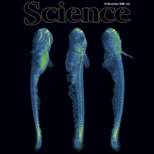
A long-standing goal of biology is to map the behavior of all cells during vertebrate embryogenesis. We developed digital scanned laser light sheet fluorescence microscopy and recorded nuclei localization and movement in entire wild-type and mutant zebrafish embryos over the first 24 hours of development. Multiview in vivo imaging at 1.5 billion voxels per minute provides "digital embryos," that is, comprehensive databases of cell positions, divisions, and migratory tracks. Our analysis of global cell division patterns reveals a maternally defined initial morphodynamic symmetry break, which identifies the embryonic body axis. We further derive a model of germ layer formation and show that the mesendoderm forms from one-third of the embryo’s cells in a single event. Our digital embryos, with 55 million nucleus entries, are provided as a resource.
Recording of the electrical activity from one of the smallest cells of a mammalian organism- a sperm cell- has been a challenging task for electrophysiologists for many decades. The method known as "spermatozoan patch clamp" was introduced in 2006. It has enabled the direct recording of ion channel activity in whole-cell and cell-attached configurations and has been instrumental in describing sperm cell physiology and the molecular identity of various calcium, potassium, sodium, chloride, and proton ion channels. However, recording from single spermatozoa requires advanced skills and training in electrophysiology. This detailed protocol summarizes the step-by-step procedure and highlights several 'tricks-of-the-trade' in order to make it available to anyone who wishes to explore the fascinating physiology of the sperm cell. Specifically, the protocol describes recording from human and murine sperm cells but can be adapted to essentially any mammalian sperm cell of any species. The protocol covers important details of the application of this technique, such as isolation of sperm cells, selection of reagents and equipment, immobilization of the highly motile cells, formation of the tight (Gigaohm) seal between a recording electrode and the plasma membrane of the sperm cells, transition into the whole-spermatozoan mode (also known as break-in), and exemplary recordings of the sperm cell calcium ion channel, CatSper, from six mammalian species. The advantages and limitations of the sperm patch clamp method, as well as the most critical steps, are discussed.