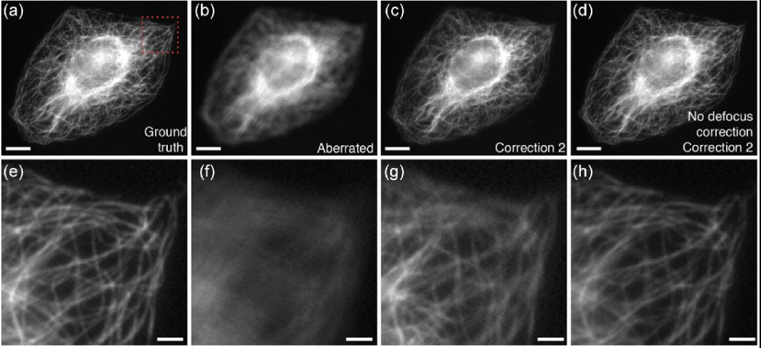
Astronomers figured out long ago how to make the images their telescopes capture of far-away galaxies clearer and sharper. By using techniques that measure how light is distorted by the atmosphere, they can apply corrections to cancel out aberrations.
Microscopists have been adapting these methods to generate clearer images of thick biological samples, which also bend light and create distortions. But these techniques – a class of methods called adaptive optics – are complex, expensive, and slow, making them out of reach for many labs.
Now, in hopes of making adaptive optics more widely available to biologists, a team led by the Shroff Lab has turned their attention to a class of techniques called phase diversity that’s been widely used in astronomy but is new to the life sciences.
These phase diversity methods add additional images with known aberrations to a blurry image with an unknown aberration, providing enough additional information to unblur the original image. Unlike many other adaptive optics techniques, phase diversity doesn’t require any major changes to an imaging system, making it a potentially attractive route for microscopy.
To implement the new method, the team first adapted the astronomy algorithm for use in microscopy and validated it with simulations. Next, they built a microscope with a deformable mirror, whose reflective surface can be changed, and two additional lenses – minor modifications to an existing microscope that create the known aberration. They also improved the software used to carry out the phase diversity correction.
As a test of their new method, the team demonstrated that they could calibrate the microscope’s deformable mirror 100 times faster than with competing methods. Next, they showed that the new method could sense and correct randomly generated aberrations, providing clearer images of fluorescent beads and fixed cells.
The next step is to test the method on real-world samples, including living cells and tissues, and extend its use to more complex microscopes. The team also hopes to make the method more automated and easier to use. They hope the new method, which is faster and cheaper to implement than current techniques, could one day make adaptive optics accessible to more labs, helping biologists see more clearly when peering deep inside tissues.
###
Citation
Courtney Johnson, Min Guo, Magdalena C. Schneider, Yijun Su, Satya Khuon, Nikolaj Reiser, Yicong Wu, Patrick La Riviere, and Hari Shroff. “Phase-diversity-based wavefront sensing for fluorescence microscopy.” Optica. Published June 10, 2024. DOI: 10.1364/OPTICA.518559
Media Contacts
Nanci Bompey


