Pinpointing which neurons process a common light-dark motion illusion could help explain how brains perceive movement

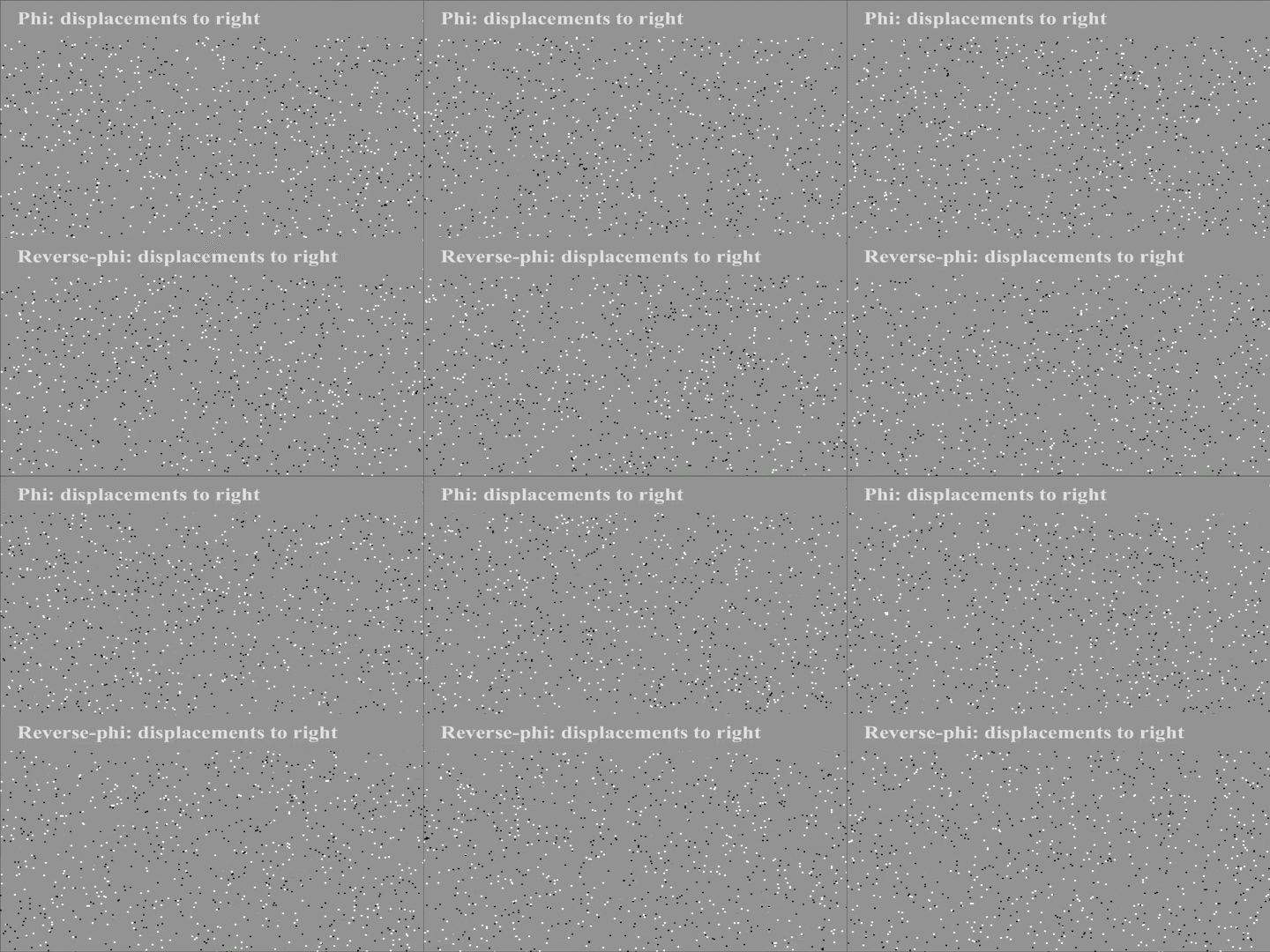
If the dots in the video look like they’re flowing in opposite directions, you’re experiencing the “reverse phi” illusion. In “phi” motion, bright points appear to move right as they disappear and reappear to the right of their previous position. But when the points switch from bright to dark as they shift rightward, our brains see them “moving” to the left: this is “reverse phi.” Now, Janelia scientist James Fitzgerald, Yale University’s Damon Clark, and their colleagues have uncovered how visual neurons in fruit flies process this illusion.
Two parallel pathways in the brain respond to either light or dark moving edges. The reverse-phi effect, like many real-world visual scenes, involves both light and dark stimuli. If the pathways segregate light from dark, the researchers asked, where in the flies’ visual system do these interacting signals combine to create a sense of motion?
Neurons called T4 and T5 cells react selectively to either light or dark moving edges. Scientists had thought that each of these cell types could respond only to light or dark stimuli. But the new results reveal that both cell types actually process a mix of light and dark signals, the team reported December 3, 2018, in the journal, Current Biology. By tracing the source of the motion illusion to the very earliest motion-detecting neurons in the fly’s visual system, the researchers showed that this light-dark mixing is a fundamental feature of the visual processing pathway.
###
Citation
Emilio Salazar-Gatzimas, Margarida Agrochao, James E. Fitzgerald, and Damon A. Clark. “The Neuronal Basis of an Illusory Motion Percept is Explained by Decorrelation of Parallel Motion Pathways.” Current Biology. Published online November 21, 2018. doi:https://doi.org/10.1016/j.cub.2018.10.007

