Main Menu (Mobile)- Block
- Overview
-
Support Teams
- Overview
- Anatomy and Histology
- Cryo-Electron Microscopy
- Electron Microscopy
- Flow Cytometry
- Gene Targeting and Transgenics
- High Performance Computing
- Immortalized Cell Line Culture
- Integrative Imaging
- Invertebrate Shared Resource
- Janelia Experimental Technology
- Mass Spectrometry
- Media Prep
- Molecular Genomics
- Primary & iPS Cell Culture
- Project Pipeline Support
- Project Technical Resources
- Quantitative Genomics
- Scientific Computing
- Viral Tools
- Vivarium
- Open Science
- You + Janelia
- About Us
Main Menu - Block
- Overview
- Anatomy and Histology
- Cryo-Electron Microscopy
- Electron Microscopy
- Flow Cytometry
- Gene Targeting and Transgenics
- High Performance Computing
- Immortalized Cell Line Culture
- Integrative Imaging
- Invertebrate Shared Resource
- Janelia Experimental Technology
- Mass Spectrometry
- Media Prep
- Molecular Genomics
- Primary & iPS Cell Culture
- Project Pipeline Support
- Project Technical Resources
- Quantitative Genomics
- Scientific Computing
- Viral Tools
- Vivarium
Complete Fly Brain Image
Complete Fly Brain Imaged at Nanoscale Resolution
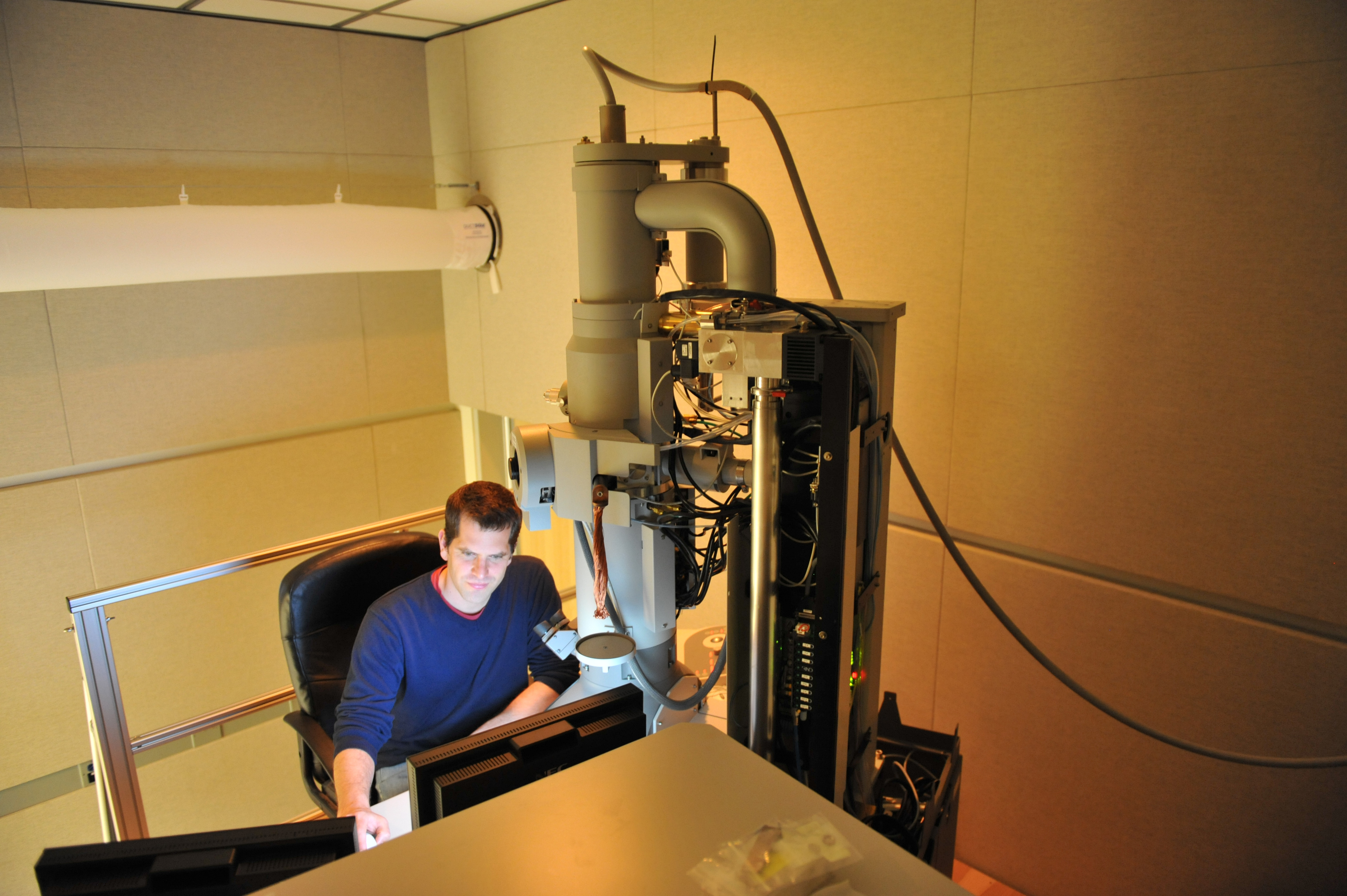
Drosophila melanogaster has a rich repertoire of innate and learned behaviors. Its 100,000-neuron brain is a large but tractable target for comprehensive neural circuit mapping. Only electron microscopy (EM) enables complete, unbiased mapping of synaptic connectivity; however, the fly brain is too large for conventional EM. Researchers led by the Bock lab developed a custom high-throughput EM platform and imaged the entire brain of an adult female fly at synaptic resolution. They traced brain-spanning circuitry involving the mushroom body (MB) to validate the dataset, which has been extensively studied for its role in learning. All inputs to Kenyon cells (KCs), the intrinsic neurons of the MB, were mapped, revealing a previously unknown cell type, postsynaptic partners of KC dendrites, and unexpected clustering of olfactory projection neurons. These reconstructions show that this freely available EM volume supports the mapping of brain-spanning circuits, which will significantly accelerate Drosophila neuroscience.
Data Description
The Bock lab at HHMI Janelia created the first-ever full mapping of all 100,000 neurons found in the brain of the Drosophila melanogaster, including Kenyon cells and their communications partners. Janelia researchers produced images by infusing the brain with a mixture of heavy metals, outlining each neuron and its connections for observation and documentation. The full data set with mapping of all neurons is available for download on the right-hand bar under a CC-BY-NC 4.0 creative commons license.


