Main Menu (Mobile)- Block
Main Menu - Block
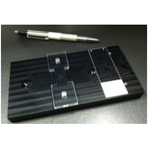
DPX Slide Jig
About the Innovation
The process of embedding tissue in the plastic resin dibutyl phthalate in xylene (DPX) creates optically transparent specimens and produces high-resolution, fluorescent confocal images. To optimize resolution and make the entire thickness accessible at high magnification, the samples are mounted directly on a coverslip. DPX is applied to the samples before the coverslip is inverted onto a prepared slide. The prepared slide supports the coverslip so that the tissue is not crushed and ensures uninhibited movement of short working distance microscope objectives.
The DPX slide jig is a simple, easy-to-use tool for quickly preparing DPX slides with exacting tolerances. The jig holds the microscope slide and the glass spacers in the correct positions, creating the optimal space for the coverslip-mounted specimens. The DPX jig is a tool that streamlines the production of uniform slides that are ideal for high-resolution imaging of DPX embedded specimens.
Advantages:
- Simplifies the preparation of DPX slides for microscopic imaging.
- Produces uniform, accurate results.
- Prevents microscope errors caused by slide components interfering with the movement of the objective.
Applications:
- Compatible with any light microscopy technique, from low-resolution widefield microscopy with air objectives to high-resolution confocal microscopy with oil immersion objectives.
- It can be adapted for mounting any tissue in any viscous mounting medium.
Opportunity:
Free to make for Non-Profit Research by downloading designs at Flintbox link to the right.
Rights and designs available for Commercial License.
For inquiries, please reference:
Janelia 2014-036