Main Menu (Mobile)- Block
- Overview
-
Support Teams
- Overview
- Anatomy and Histology
- Cryo-Electron Microscopy
- Electron Microscopy
- Flow Cytometry
- Gene Targeting and Transgenics
- High Performance Computing
- Immortalized Cell Line Culture
- Integrative Imaging
- Invertebrate Shared Resource
- Janelia Experimental Technology
- Mass Spectrometry
- Media Prep
- Molecular Genomics
- Stem Cell & Primary Culture
- Project Pipeline Support
- Project Technical Resources
- Quantitative Genomics
- Scientific Computing
- Viral Tools
- Vivarium
- Open Science
- You + Janelia
- About Us
Main Menu - Block
- Overview
- Anatomy and Histology
- Cryo-Electron Microscopy
- Electron Microscopy
- Flow Cytometry
- Gene Targeting and Transgenics
- High Performance Computing
- Immortalized Cell Line Culture
- Integrative Imaging
- Invertebrate Shared Resource
- Janelia Experimental Technology
- Mass Spectrometry
- Media Prep
- Molecular Genomics
- Stem Cell & Primary Culture
- Project Pipeline Support
- Project Technical Resources
- Quantitative Genomics
- Scientific Computing
- Viral Tools
- Vivarium
Enhanced FIB-SEM
Enhanced Focused Ion Beam Scanning Electron Microscopy (FIB-SEM)
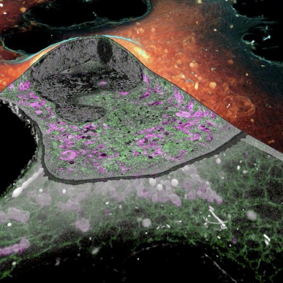
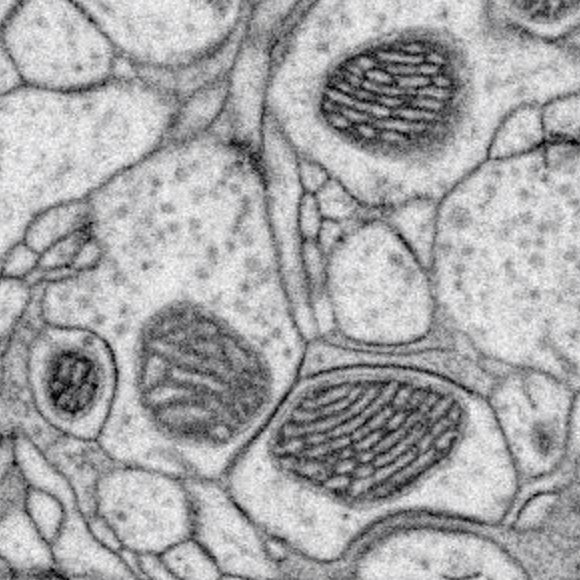
Focused Ion Beam Scanning Electron Microscopy (FIB-SEM) is an advanced technique to make 3D images of biological cells in tissues with superior z-axis (vertical) resolution, generating easily interpretable data for reconstruction. FIB-SEM produces detailed images of cell features like protein complexes and membranes through an electron beam's sequential etching of a sample surface. Until the developments of Enhanced FIB-SEM by C. Shan Xu and Harald Hess, presented here, the image produced had been limited to a single cell or small volume of cells.
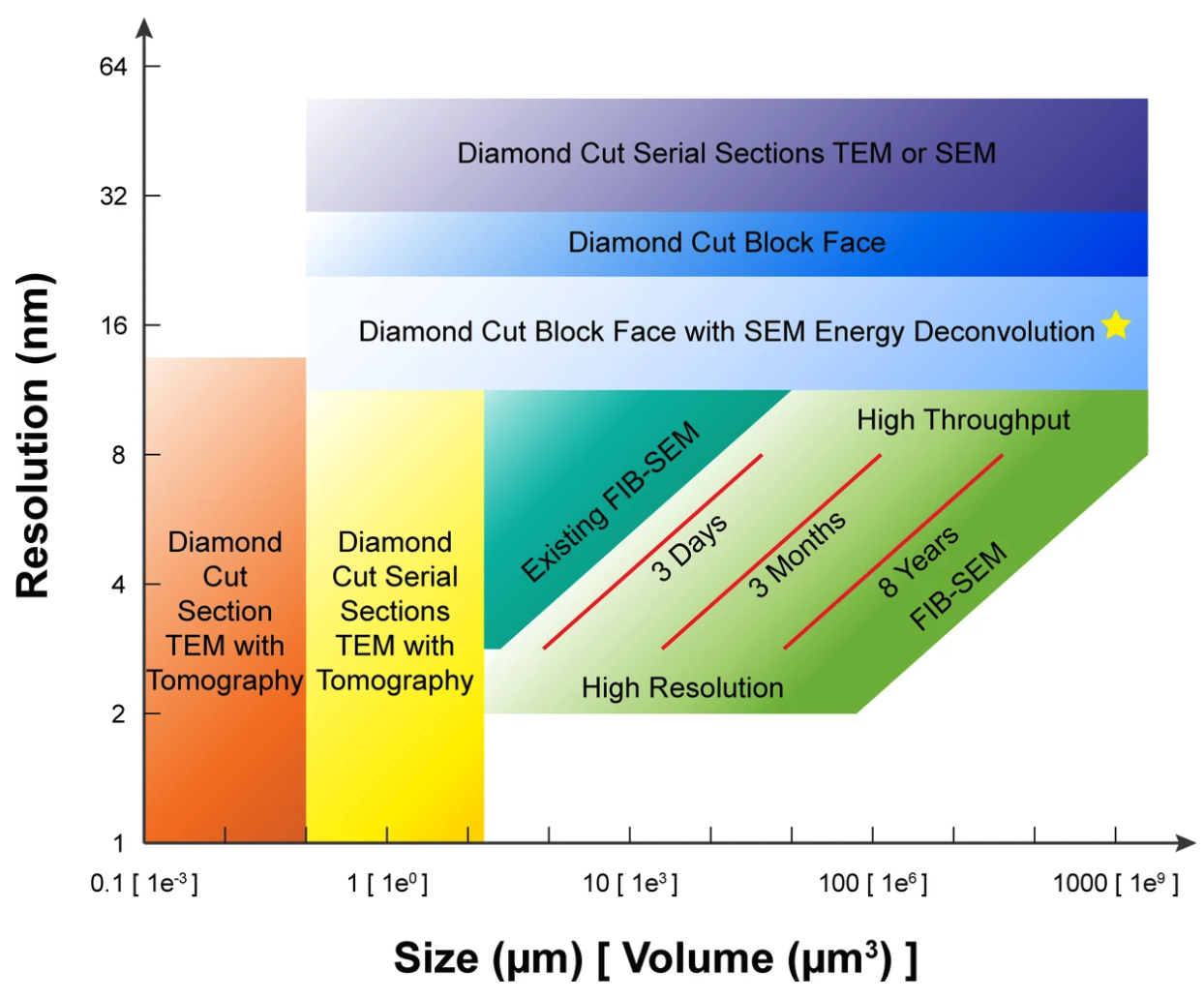
The inherent technical limitations on FIB-SEM were slow imaging speed and lack of long-term system stability, which improved the abilities of TEM, still limited the image volume. However, the Hess lab masterfully re-engineered the FIB-SEM instrument orientation, reliability, and self-correction components, developed instrument designs, and techniques to accelerate image acquisition, and achieving dramatically improved performance.
This development continues with the full resources and attention of the FIB-SEM Technology, led by C. Shan Xu.
The enhanced FIB-SEM methods allow a system to operate continuously for months and image volumes greater than 106 µm3. At this range, connectomics studies and mapping neuronal processes is possible, and it avoids tedious human proofreading.
The subject of an issued US Patent, enhanced FIB-SEM includes the following features:
-
In-line automatic focus, stigmation, and aperture alignment optimization
-
Temperature and currents monitoring
-
Closed-loop control of FIB milling
-
Email notification for warnings and auto-pause of operation to avoid sample damage
-
Auto restart after a pause
-
Multiple magnifications at user-selected intervals
-
Real-time image alignment
In more detail in the publication, the high-impact developments described here have made a substantial change in FIB-SEM enabled research, as shown by the 145 citations times of the first publication. The patent rights and designs are available for a commercial license for manufacture and sale.
Intellectual Property:
US Patent 10,600,615
European Patent Application EP3574518A2
Inventors:
C. Shan Xu
Kenneth Jeffrey Hayworth
Harald Hess
Tech ID: 2014-017


