Main Menu (Mobile)- Block
- Overview
-
Support Teams
- Overview
- Anatomy and Histology
- Cryo-Electron Microscopy
- Electron Microscopy
- Flow Cytometry
- Gene Targeting and Transgenics
- High Performance Computing
- Immortalized Cell Line Culture
- Integrative Imaging
- Invertebrate Shared Resource
- Janelia Experimental Technology
- Mass Spectrometry
- Media Prep
- Molecular Genomics
- Primary & iPS Cell Culture
- Project Pipeline Support
- Project Technical Resources
- Quantitative Genomics
- Scientific Computing
- Viral Tools
- Vivarium
- Open Science
- You + Janelia
- About Us
Main Menu - Block
- Overview
- Anatomy and Histology
- Cryo-Electron Microscopy
- Electron Microscopy
- Flow Cytometry
- Gene Targeting and Transgenics
- High Performance Computing
- Immortalized Cell Line Culture
- Integrative Imaging
- Invertebrate Shared Resource
- Janelia Experimental Technology
- Mass Spectrometry
- Media Prep
- Molecular Genomics
- Primary & iPS Cell Culture
- Project Pipeline Support
- Project Technical Resources
- Quantitative Genomics
- Scientific Computing
- Viral Tools
- Vivarium
Oscillating Stimulus Transporter Assay
A simplified, high-throughput-capable assay for measuring transporter function
Membrane-bound transporter proteins enable passage of nearly all biologically-relevant molecules across lipid membranes. They are critical for cell nutrition and communication, and are targeted by innumerable drug classes.
Study of transporters is limited by the difficulty in handling them outside of a membrane and experimental protocols that are laborious, need dedicated, specialized equipment, and have extensive controls. Applying simple fluorescence techniques to these experiments would lead to better, easier experiments.
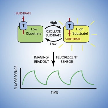
Researchers at HHMI's Janelia Research Campus have developed a novel assay to enable rapid and easy measurement of many transporter properties. The setup is simple: cells expressing a transporter of interest are loaded with small-molecule or genetically-encoded fluorescent sensors. Then the cells are perfused with solutions oscillating between high and low substrate concentrations. Optical detection is analyzed with free software such as ImageJ/FIJI and transporter properties are characterized from the signals.
The researchers have demonstrated transporter analyses with this technique on a glutamate transporter, Cl-/HCO3- exchangers, Na+/Glucose co-transporters, Glucose transporters and others. In nearly physiological conditions, measurements of inhibition, mobilization, and transport rates can be easily accessed. A setup may involve basic, available equipment or may be improved through advanced instrumentation in development. Basic requirement are cells of interest, plasmids for transporter and reporter, and a method for measuring time-dependent fluorescence (usually fluorescence microscopic imaging.) Further development may enable surface plasmon resonance- (SPR-) type measurements of pharmacological binding kinetics.
Advantages:
- Rapidly characterizes transporters with a standard equipment
- Uses near-physiological conditions
- Uses high signal-to-noise fluorescence detection
- Simple experimental setup: transfect and measure
- Analysis software widely available
Applications:
- Transporter characterization
- Drug screening
- Rapidly assess activity and KD’s of inhibition for drugs or other compounds
- Potentially assess kinetics of binding/inhibition for natural agents
Opportunity:
Commercial License or Non-Profit Research
For inquiries, please reference:
Janelia 2016-043

