Main Menu (Mobile)- Block
- Overview
-
Support Teams
- Overview
- Anatomy and Histology
- Cryo-Electron Microscopy
- Electron Microscopy
- Flow Cytometry
- Gene Targeting and Transgenics
- High Performance Computing
- Immortalized Cell Line Culture
- Integrative Imaging
- Invertebrate Shared Resource
- Janelia Experimental Technology
- Mass Spectrometry
- Media Prep
- Molecular Genomics
- Stem Cell & Primary Culture
- Project Pipeline Support
- Project Technical Resources
- Quantitative Genomics
- Scientific Computing
- Viral Tools
- Vivarium
- Open Science
- You + Janelia
- About Us
Main Menu - Block
- Overview
- Anatomy and Histology
- Cryo-Electron Microscopy
- Electron Microscopy
- Flow Cytometry
- Gene Targeting and Transgenics
- High Performance Computing
- Immortalized Cell Line Culture
- Integrative Imaging
- Invertebrate Shared Resource
- Janelia Experimental Technology
- Mass Spectrometry
- Media Prep
- Molecular Genomics
- Stem Cell & Primary Culture
- Project Pipeline Support
- Project Technical Resources
- Quantitative Genomics
- Scientific Computing
- Viral Tools
- Vivarium
Vibratome built for the Auto Slicer Imager Rig (MouseLight 2)
About the Innovation
Visualization of the axonal structures of individual neurons is critical to understand how neural signals are organized and communicated in the brain. Janelia researchers developed an imaging system for whole-brain, high-resolution fluorescence imaging. They showed that researchers could use it to trace individual axonal fibers and fine axon collaterals across the brain. These can map the long-distance projections of single neurons.
The technology offered here is a platform for high-resolution, three-dimensional fluorescence imaging of complete tissue volumes. They enable visualization and reconstruction of long-range axonal arbors. It uses a high-speed two-photon microscope with a tissue vibratome made from a modified Leica Vibratome and a suite of computational tools for large-scale image data.
The related publication (https://elifesciences.org/articles/10566) shows the power of this approach by reconstructing the axonal arbors of multiple neurons in the motor cortex across a single mouse brain.
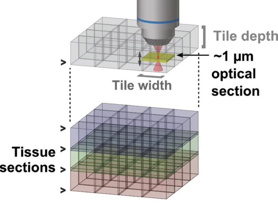
The system is fully automated and made with commercially available hardware, including a mounting block for the vibratome, other custom parts, and custom control software. It works by imaging a layer of tissue near the exposed surface of a sample then cutting off a slice of that imaged tissue volume. The steps repeat until the entire sample has been imaged; this takes about a week for a whole mouse brain and produces about 30 terabytes of images.
Linked here are a compilation/CAD drawings of custom parts needed to mount a modified Leica 1200 vibratome on a 2-photon microscope that does automated continuous volumetric imaging of an entire mouse brain.
Opportunity
Free to make for Non-Profit Research by downloading designs at Flintbox link to the right.
Rights and designs available for Commercial License.
For inquiries, please reference:
Janelia 2017-051


