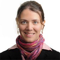
Current Research:
My current research is focused on protein engineering molecular tools in order to make microscopy better. Currently my main focus is fixation resistant molecular tools. By changing surface-reactive amino acids one can manipulate the protein to be resistant to fixation, while maintaining its protein functions.
Biography
I am a research specialist in the Looger lab. I have a deep love for protein biochemistry, and molecular biology. I grew up in San Luis-Argentina, where I got my bachelors in biochemistry in 2004, following my Dad’s footsteps, in the National University of San Luis-Argentina. During my schooling I also worked as a research technician in the Organic Chemistry department of the same university (2001-2004). At this point, I became the first person in my entire family to leave my home country when I traveled to the USA. I enrolled in the Chemistry and Biochemistry department of the University of Milwaukee-Wisconsin, where I did research on two projects. The first project was with Dr. Guillerme Indig (Chemistry department), where I acquired most of my light and fluorescence microscopy knowledge, and cancer cell development. Eventually part of my work was published in a paper called “Mitochondrial targeting for photochemotherapy: Can selective tumor cell killing be predicted based on n-octanol/water distribution coefficients?" (in Biotechnique & Histochemistry). The second project was with Professor Steven Forst (Biology department), where I acquired most of my knowledge about molecular biology. The name of my M.S. project was “Cloning, expression, purification, and biochemical analysis of Var-1: a novel protein of Xenorhabdus nematophila” (paper in preparation). Soon after I completed my M.S. degree in Biochemistry & Chemistry (Aug. 2008), I relocated to Washington DC, after my husband got a job at the National Science Foundation. A few months later, I was hired by Dr. Doug Murphy at Janelia Research Campus to temporarily work at the microscopy facility for eight months (four months at the microscopy facility followed by four months at cell culture facility). During that time I assisted more than 20 postdoctoral fellows with experiments in microscopy. Specifically I helped them to acquire, process, save in different formats, and analyze images using fluorescent or light microscopy. The instruments that I was using were 4 confocal systems (one with 2-photon incorporated), and 3 wide-field systems, for either live-cell imaging or fixed specimens, using different cell cultures. Specifically Heck 293 (human kidney cells transformed with adenovirus 5); HeLa cells (human epithelial cells from cervix adenocarcinoma); adeno-associated virus cell line (derived from HEK 293 cell line). Furthermore I transfected them with different protocols (including, Polyplus, FuGENE HD, and Amaxa). In September of 2010 Dr. Loren Looger permanently hired me where I have handled several projects, as well as having the opportunity to work on my own assigned project (mEos4 project). Currently (Sept. 2011) I work in the designing and engineering of new molecules and protocols for CLEM (Correlative Light and Electron Microscopy). In particular, I have made substantial progress in engineering a version of mEos with increased protein stability (mEos2 to mEos4). mEos2 was the last fluorescent protein designed by Sean McKinney in the Looger lab. It is a photoconvertible fluorescent protein (changes fluorescence from green to red by hitting with UV light), and basically is the best standard photoconvertible FP (fluorescent protein) until date. Our codename for a better mEos2 now is mEos4. I first began by working on improving the thermal stability, followed by extensive work on fixatives and auto-fluorescence of different variants of mEos4. With the electron microscopy (EM) team, Richard Fetter, Yalin Wang, and Mei Sun, our main discovery has been a new protocol for EM that will allow us to use CLEM for any fluorophore and fluorescent protein, by decreasing the auto-fluorescence of fixatives, the harsh conditions of OsO4, and increasing the ultra-structure preservation of cell tissue ( http://www.nature.com/nmeth/journal/v12/n3/full/nmeth.3225.html). Specifically, I am working on improving horseradish peroxidase (HRP), psCFP2, and other fluorescent proteins to fixation.
Education
Previous Experience
Microscopy and cell culture assistant.
I have assisted postdoctoral fellows with experiments in microscopy (Specifically I helped them to acquire, process, save in different formats, and analyze images using fluorescent or light microscopy. The instruments that I was using were 4 confocal systems (one with 2-photon incorporated), and 3 wide-field systems, for either live-cell imaging or fixed specimens, cell culture [different culture of cells. Specifically 293[Heck 293] (human kidney cells transformed with adenovirus 5); HeLa cells [CCL-2](human epithelial cells from cervix adenocarcinoma); Adeno-associated virus cell line (derived from HEK 293 cell line) for post-docs, or PIs. Furthermore I transfect them with different techniques. Specifically Polyplus, and FuGENE HD, Amaxa] and protein biochemistry (DNA constructs, protein purification, and characterization).
Teaching assistant.
Assisted my Instructor-professor when needed in teaching students for laboratory practice in chemistry and biochemistry. Prepare all the necessary instruments for the lab. such as glass, and drugs. I performed tutoring; holding office hours; tests or exams; and assisted a professor with a large lecture class by teaching students in recitation, laboratory, or discussion sessions. Furthermore, I assisted to help teach discussions during regular class.
