Main Menu (Mobile)- Block
Main Menu - Block


Abstract
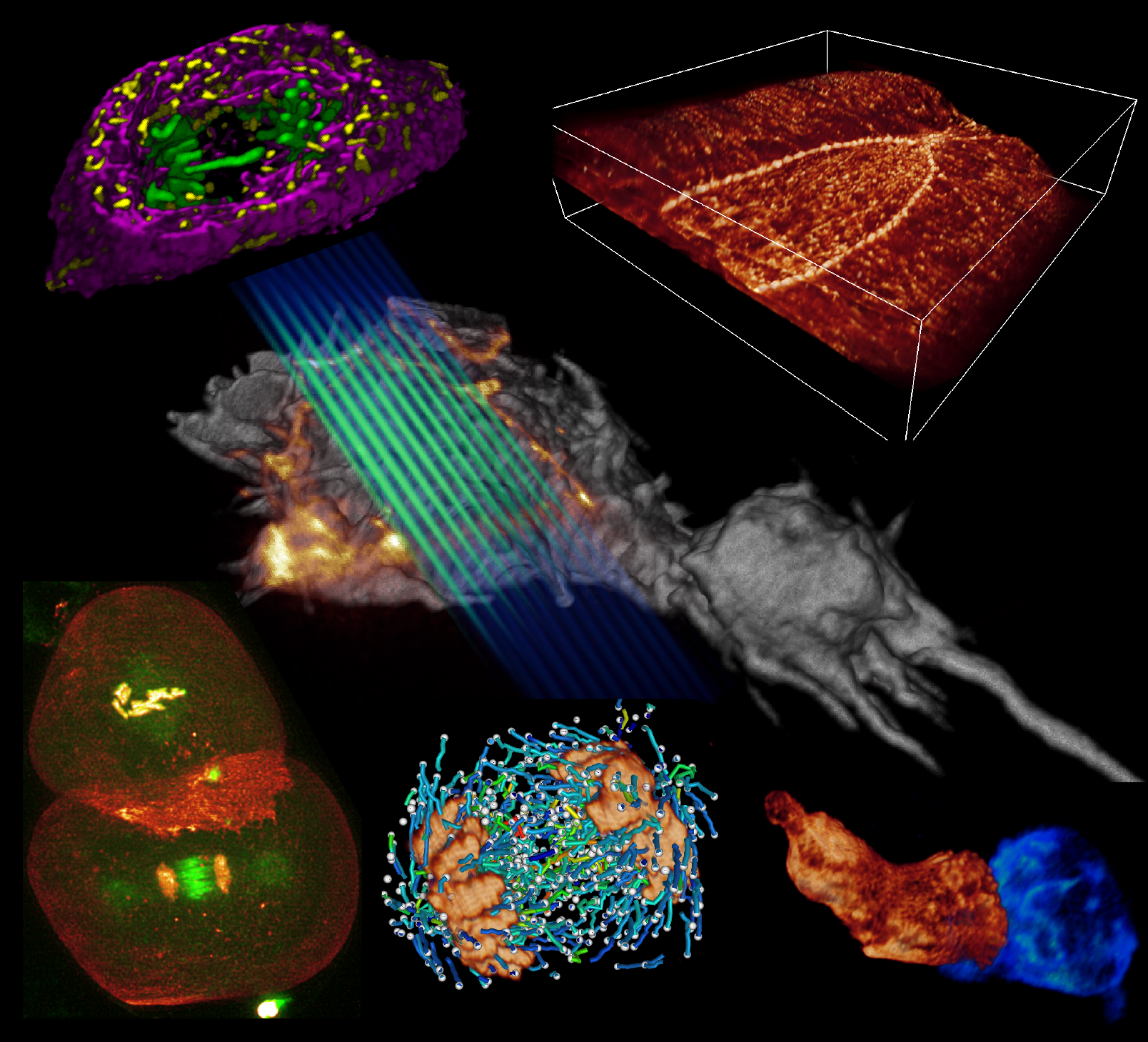
A key challenge when imaging living cells is how to noninvasively extract the most spatiotemporal information possible. Unlike popular wide-field and confocal methods, plane-illumination microscopy limits excitation to the information-rich vicinity of the focal plane, providing effective optical sectioning and high speed while minimizing out-of-focus background and premature photobleaching. Here we used scanned Bessel beams in conjunction with structured illumination and/or two-photon excitation to create thinner light sheets (<0.5 μm) better suited to three-dimensional (3D) subcellular imaging. As demonstrated by imaging the dynamics of mitochondria, filopodia, membrane ruffles, intracellular vesicles and mitotic chromosomes in live cells, the microscope currently offers 3D isotropic resolution down to \~{}0.3 μm, speeds up to nearly 200 image planes per second and the ability to noninvasively acquire hundreds of 3D data volumes from single living cells encompassing tens of thousands of image frames.
Commentary: Plane illumination microscopy has proven to be a powerful tool for studying multicellular organisms and their development at single cell resolution. However, the light sheets employed are usually too thick to provide much benefit for imaging organelles within single cultured cells. Here we introduce the use of scanned Bessel beams to create much thinner light sheets better suited to long-term dynamic live cell imaging. Such light sheets not only minimize photobleaching and phototoxicity at the sub-cellular level, but also provide axial resolution enhancement, yielding isotropic three dimensional spatial resolution. Numerous movies are provided to demonstrate the wealth of 4D information (x,y,x,t) that can be obtained from single living cells by the method. Besides providing an attractive alternative to spinning disk, AOD-driven, or line scan confocal microscopes for high speed live cell imaging, the Bessel microscope might serve as a valuable platform for superresolution microscopy (PALM, structured Illumination, or RESOLFT), since confinement of the excitation to the focal plane makes far better use of the limited fluorescence photon budget than does the traditional epi-illumination configuration.