Filter
Associated Lab
- Ahrens Lab (2) Apply Ahrens Lab filter
- Betzig Lab (9) Apply Betzig Lab filter
- Hess Lab (1) Apply Hess Lab filter
- Ji Lab (2) Apply Ji Lab filter
- Lavis Lab (1) Apply Lavis Lab filter
- Pedram Lab (2) Apply Pedram Lab filter
- Saalfeld Lab (1) Apply Saalfeld Lab filter
- Remove Shroff Lab filter Shroff Lab
- Wang (Shaohe) Lab (2) Apply Wang (Shaohe) Lab filter
Associated Project Team
Associated Support Team
31 Janelia Publications
Showing 1-10 of 31 resultsNeurobiological processes occur on spatiotemporal scales spanning many orders of magnitude. Greater understanding of these processes therefore demands improvements in the tools used in their study. Here we review recent efforts to enhance the speed and resolution of one such tool, fluorescence microscopy, with an eye toward its application to neurobiological problems. On the speed front, improvements in beam scanning technology, signal generation rates, and photodamage mediation are bringing us closer to the goal of real-time functional imaging of extended neural networks. With regard to resolution, emerging methods of adaptive optics may lead to diffraction-limited imaging or much deeper imaging in optically inhomogeneous tissues, and super-resolution techniques may prove a powerful adjunct to electron microscopic methods for nanometric neural circuit reconstruction.
Neurobiological processes occur on spatiotemporal scales spanning many orders of magnitude. Greater understanding of these processes therefore demands improvements in the tools used in their study. Here we review recent efforts to enhance the speed and resolution of one such tool, fluorescence microscopy, with an eye toward its application to neurobiological problems. On the speed front, improvements in beam scanning technology, signal generation rates, and photodamage mediation are bringing us closer to the goal of real-time functional imaging of extended neural networks. With regard to resolution, emerging methods of adaptive optics may lead to diffraction-limited imaging or much deeper imaging in optically inhomogeneous tissues, and super-resolution techniques may prove a powerful adjunct to electron microscopic methods for nanometric neural circuit reconstruction.
Commentary: A brief review of recent trends in microscopy. The section “Caveats regarding the application of superresolution microscopy” was written in an effort to inject a dose of reality and caution into the unquestioning enthusiasm in the academic community for all things superresolution, covering the topics of labeling density and specificity, sample preparation artifacts, speed vs. resolution vs. photodamage, and the implications of signal-to-background for Nyquist vs. Rayleigh definitions of resolution.
Starvation triggers bacterial spore formation, a committed differentiation program that transforms a vegetative cell into a dormant spore. Cells in a population enter sporulation nonuniformly to secure against the possibility that favorable growth conditions, which put sporulation-committed cells at a disadvantage, may resume. This heterogeneous behavior is initiated by a passive mechanism: stochastic activation of a master transcriptional regulator. Here, we identify a cell-cell communication pathway containing the proteins ShfA (YabQ) and ShfP (YvnB) that actively promotes phenotypic heterogeneity, wherein Bacillus subtilis cells that start sporulating early use a calcineurin-like phosphoesterase to release glycerol, which simultaneously acts as a signaling molecule and a nutrient to delay nonsporulating cells from entering sporulation. This produced a more diverse population that was better poised to exploit a sudden influx of nutrients compared to those generating heterogeneity via stochastic gene expression alone. Although conflict systems are prevalent among microbes, genetically encoded cooperative behavior in unicellular organisms can evidently also boost inclusive fitness.
The intestine is critical for not only processing nutrients but also protecting the organism from the environment. These functions are mainly carried out by the epithelium, which is constantly being self-renewed. Many genes and pathways can influence intestinal epithelial cell proliferation. Among them is mTORC1, whose activation increases cell proliferation. Here, we report the first intestinal epithelial cell (IEC)-specific knockout () of an amino acid transporter capable of activating mTORC1. We show that the transporter, SLC7A5, is highly expressed in mouse intestinal crypt and reduces mTORC1 signaling. Surprisingly, adult intestinal crypts have increased cell proliferation but reduced mature Paneth cells. Goblet cells, the other major secretory cell type in the small intestine, are increased in the crypts but reduced in the villi. Analyses with scRNA-seq and electron microscopy have revealed dedifferentiation of Paneth cells in mice, leading to markedly reduced secretory granules with little effect on Paneth cell number. Thus, SLC7A5 likely regulates secretory cell differentiation to affect stem cell niche and indirectly regulate cell proliferation.
When faced with starvation, the bacterium transforms itself into a dormant cell type called a "spore". Sporulation initiates with an asymmetric division event, which requires the relocation of the core divisome components FtsA and FtsZ, after which the sigma factor σ is exclusively activated in the smaller daughter cell. Compartment-specific activation of σ requires the SpoIIE phosphatase, which displays a biased localization on one side of the asymmetric division septum and associates with the structural protein DivIVA, but the mechanism by which this preferential localization is achieved is unclear. Here, we isolated a variant of DivIVA that indiscriminately activates σ in both daughter cells due to promiscuous localization of SpoIIE, which was corrected by overproduction of FtsA and FtsZ. We propose that the core components of the redeployed cell division machinery drive the asymmetric localization of DivIVA and SpoIIE to trigger the initiation of the sporulation program.
Deep neural networks have been applied to improve the image quality of fluorescence microscopy imaging. Previous methods are based on convolutional neural networks (CNNs) which generally require more time-consuming training of separate models for each new imaging experiment, impairing the applicability and generalization. Once the model is trained (typically with tens to hundreds of image pairs) it can then be used to enhance new images that are like the training data. In this study, we proposed a novel imaging-transformer based model, Convolutional Neural Network Transformer (CNNT), to outperform the CNN networks for image denoising. In our scheme we have trained a single CNNT based backbone model from pairwise high-low SNR images for one type of fluorescence microscope (instance structured illumination, iSim). Fast adaption to new applications was achieved by fine-tuning the backbone on only 5-10 sample pairs per new experiment. Results show the CNNT backbone and fine-tuning scheme significantly reduces the training time and improves the image quality, outperformed training separate models using CNN approaches such as - RCAN and Noise2Fast. Here we show three examples of the efficacy of this approach on denoising wide-field, two-photon and confocal fluorescence data. In the confocal experiment, which is a 5 by 5 tiled acquisition, the fine-tuned CNNT model reduces the scan time form one hour to eight minutes, with improved quality.
Deep neural networks can improve the quality of fluorescence microscopy images. Previous methods, based on Convolutional Neural Networks (CNNs), require time-consuming training of individual models for each experiment, impairing their applicability and generalization. In this study, we propose a novel imaging-transformer based model, Convolutional Neural Network Transformer (CNNT), that outperforms CNN based networks for image denoising. We train a general CNNT based backbone model from pairwise high-low Signal-to-Noise Ratio (SNR) image volumes, gathered from a single type of fluorescence microscope, an instant Structured Illumination Microscope. Fast adaptation to new microscopes is achieved by fine-tuning the backbone on only 5-10 image volume pairs per new experiment. Results show that the CNNT backbone and fine-tuning scheme significantly reduces training time and improves image quality, outperforming models trained using only CNNs such as 3D-RCAN and Noise2Fast. We show three examples of efficacy of this approach in wide-field, two-photon, and confocal fluorescence microscopy.
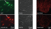
Optical aberrations hinder fluorescence microscopy of thick samples, reducing image signal, contrast, and resolution. Here we introduce a deep learning-based strategy for aberration compensation, improving image quality without slowing image acquisition, applying additional dose, or introducing more optics. Our method (i) introduces synthetic aberrations to images acquired on the shallow side of image stacks, making them resemble those acquired deeper into the volume and (ii) trains neural networks to reverse the effect of these aberrations. We use simulations and experiments to show that applying the trained ‘de-aberration’ networks outperforms alternative methods, providing restoration on par with adaptive optics techniques; and subsequently apply the networks to diverse datasets captured with confocal, light-sheet, multi-photon, and super-resolution microscopy. In all cases, the improved quality of the restored data facilitates qualitative image inspection and improves downstream image quantitation, including orientational analysis of blood vessels in mouse tissue and improved membrane and nuclear segmentation in C. elegans embryos.
Sample-induced aberrations and optical imperfections limit the resolution of fluorescence microscopy. Phase diversity is a powerful technique that leverages complementary phase information in sequentially acquired images with deliberately introduced aberrations--the phase diversities--to enable phase and object reconstruction and restore diffraction-limited resolution. These phase diversities are typically introduced into the optical path via a deformable mirror. Existing phase-diversity-based methods are limited to Zernike modes, require large numbers of diversity images, or depend on accurate mirror calibration--which are all suboptimal. We present DeepPD, a deep learning-based framework that combines neural representations of the object and wavefront with a learned model of the deformable mirror to jointly estimate both object and phase from only five images. DeepPD improves robustness and reconstruction quality over previous approaches, even under severe aberrations. We demonstrate its performance on calibration targets and biological samples, including immunolabeled myosin in fixed PtK2 cells.
Germ granules, specialized ribonucleoprotein particles, are a hallmark of all germ cells. In Drosophila, an estimated 200 mRNAs are enriched in the germ plasm, and some of these have important, often conserved roles in germ cell formation, specification, survival and migration. How mRNAs are spatially distributed within a germ granule and whether their position defines functional properties is unclear. Here we show, using single-molecule FISH and structured illumination microscopy, a super-resolution approach, that mRNAs are spatially organized within the granule whereas core germ plasm proteins are distributed evenly throughout the granule. Multiple copies of single mRNAs organize into 'homotypic clusters' that occupy defined positions within the center or periphery of the granule. This organization, which is maintained during embryogenesis and independent of the translational or degradation activity of mRNAs, reveals new regulatory mechanisms for germ plasm mRNAs that may be applicable to other mRNA granules.