Filter
Associated Lab
- Aguilera Castrejon Lab (4) Apply Aguilera Castrejon Lab filter
- Ahrens Lab (64) Apply Ahrens Lab filter
- Aso Lab (42) Apply Aso Lab filter
- Baker Lab (19) Apply Baker Lab filter
- Betzig Lab (104) Apply Betzig Lab filter
- Beyene Lab (10) Apply Beyene Lab filter
- Bock Lab (14) Apply Bock Lab filter
- Branson Lab (53) Apply Branson Lab filter
- Card Lab (37) Apply Card Lab filter
- Cardona Lab (45) Apply Cardona Lab filter
- Chklovskii Lab (10) Apply Chklovskii Lab filter
- Clapham Lab (15) Apply Clapham Lab filter
- Cui Lab (19) Apply Cui Lab filter
- Darshan Lab (8) Apply Darshan Lab filter
- Dennis Lab (2) Apply Dennis Lab filter
- Dickson Lab (32) Apply Dickson Lab filter
- Druckmann Lab (21) Apply Druckmann Lab filter
- Dudman Lab (46) Apply Dudman Lab filter
- Eddy/Rivas Lab (30) Apply Eddy/Rivas Lab filter
- Egnor Lab (4) Apply Egnor Lab filter
- Espinosa Medina Lab (21) Apply Espinosa Medina Lab filter
- Feliciano Lab (16) Apply Feliciano Lab filter
- Fetter Lab (31) Apply Fetter Lab filter
- FIB-SEM Technology (1) Apply FIB-SEM Technology filter
- Fitzgerald Lab (17) Apply Fitzgerald Lab filter
- Freeman Lab (15) Apply Freeman Lab filter
- Funke Lab (46) Apply Funke Lab filter
- Gonen Lab (59) Apply Gonen Lab filter
- Grigorieff Lab (34) Apply Grigorieff Lab filter
- Harris Lab (55) Apply Harris Lab filter
- Heberlein Lab (13) Apply Heberlein Lab filter
- Hermundstad Lab (28) Apply Hermundstad Lab filter
- Hess Lab (77) Apply Hess Lab filter
- Ilanges Lab (4) Apply Ilanges Lab filter
- Jayaraman Lab (45) Apply Jayaraman Lab filter
- Ji Lab (33) Apply Ji Lab filter
- Johnson Lab (2) Apply Johnson Lab filter
- Karpova Lab (14) Apply Karpova Lab filter
- Keleman Lab (8) Apply Keleman Lab filter
- Keller Lab (62) Apply Keller Lab filter
- Koay Lab (4) Apply Koay Lab filter
- Lavis Lab (150) Apply Lavis Lab filter
- Lee (Albert) Lab (29) Apply Lee (Albert) Lab filter
- Leonardo Lab (19) Apply Leonardo Lab filter
- Li Lab (8) Apply Li Lab filter
- Lippincott-Schwartz Lab (110) Apply Lippincott-Schwartz Lab filter
- Liu (Yin) Lab (4) Apply Liu (Yin) Lab filter
- Liu (Zhe) Lab (60) Apply Liu (Zhe) Lab filter
- Looger Lab (137) Apply Looger Lab filter
- Magee Lab (31) Apply Magee Lab filter
- Menon Lab (12) Apply Menon Lab filter
- Murphy Lab (6) Apply Murphy Lab filter
- O'Shea Lab (7) Apply O'Shea Lab filter
- Otopalik Lab (1) Apply Otopalik Lab filter
- Pachitariu Lab (44) Apply Pachitariu Lab filter
- Pastalkova Lab (6) Apply Pastalkova Lab filter
- Pavlopoulos Lab (7) Apply Pavlopoulos Lab filter
- Pedram Lab (4) Apply Pedram Lab filter
- Podgorski Lab (16) Apply Podgorski Lab filter
- Reiser Lab (49) Apply Reiser Lab filter
- Riddiford Lab (20) Apply Riddiford Lab filter
- Romani Lab (40) Apply Romani Lab filter
- Rubin Lab (111) Apply Rubin Lab filter
- Saalfeld Lab (49) Apply Saalfeld Lab filter
- Satou Lab (3) Apply Satou Lab filter
- Scheffer Lab (38) Apply Scheffer Lab filter
- Schreiter Lab (55) Apply Schreiter Lab filter
- Schulze Lab (1) Apply Schulze Lab filter
- Sgro Lab (3) Apply Sgro Lab filter
- Shroff Lab (35) Apply Shroff Lab filter
- Simpson Lab (18) Apply Simpson Lab filter
- Singer Lab (37) Apply Singer Lab filter
- Spruston Lab (62) Apply Spruston Lab filter
- Stern Lab (77) Apply Stern Lab filter
- Sternson Lab (47) Apply Sternson Lab filter
- Stringer Lab (41) Apply Stringer Lab filter
- Svoboda Lab (132) Apply Svoboda Lab filter
- Tebo Lab (12) Apply Tebo Lab filter
- Tervo Lab (10) Apply Tervo Lab filter
- Tillberg Lab (19) Apply Tillberg Lab filter
- Tjian Lab (17) Apply Tjian Lab filter
- Truman Lab (58) Apply Truman Lab filter
- Turaga Lab (41) Apply Turaga Lab filter
- Turner Lab (27) Apply Turner Lab filter
- Vale Lab (8) Apply Vale Lab filter
- Voigts Lab (5) Apply Voigts Lab filter
- Wang (Meng) Lab (31) Apply Wang (Meng) Lab filter
- Wang (Shaohe) Lab (6) Apply Wang (Shaohe) Lab filter
- Wong-Campos Lab (1) Apply Wong-Campos Lab filter
- Wu Lab (8) Apply Wu Lab filter
- Zlatic Lab (26) Apply Zlatic Lab filter
- Zuker Lab (5) Apply Zuker Lab filter
Associated Project Team
- CellMap (13) Apply CellMap filter
- COSEM (3) Apply COSEM filter
- FIB-SEM Technology (5) Apply FIB-SEM Technology filter
- Fly Descending Interneuron (12) Apply Fly Descending Interneuron filter
- Fly Functional Connectome (14) Apply Fly Functional Connectome filter
- Fly Olympiad (5) Apply Fly Olympiad filter
- FlyEM (56) Apply FlyEM filter
- FlyLight (50) Apply FlyLight filter
- GENIE (47) Apply GENIE filter
- Integrative Imaging (11) Apply Integrative Imaging filter
- Larval Olympiad (2) Apply Larval Olympiad filter
- MouseLight (18) Apply MouseLight filter
- NeuroSeq (1) Apply NeuroSeq filter
- ThalamoSeq (1) Apply ThalamoSeq filter
- Tool Translation Team (T3) (29) Apply Tool Translation Team (T3) filter
- Transcription Imaging (45) Apply Transcription Imaging filter
Associated Support Team
- Project Pipeline Support (5) Apply Project Pipeline Support filter
- Anatomy and Histology (18) Apply Anatomy and Histology filter
- Cryo-Electron Microscopy (49) Apply Cryo-Electron Microscopy filter
- Electron Microscopy (18) Apply Electron Microscopy filter
- Gene Targeting and Transgenics (11) Apply Gene Targeting and Transgenics filter
- High Performance Computing (7) Apply High Performance Computing filter
- Integrative Imaging (25) Apply Integrative Imaging filter
- Invertebrate Shared Resource (40) Apply Invertebrate Shared Resource filter
- Janelia Experimental Technology (40) Apply Janelia Experimental Technology filter
- Management Team (1) Apply Management Team filter
- Mass Spectrometry (1) Apply Mass Spectrometry filter
- Molecular Genomics (15) Apply Molecular Genomics filter
- Project Technical Resources (54) Apply Project Technical Resources filter
- Quantitative Genomics (20) Apply Quantitative Genomics filter
- Scientific Computing (106) Apply Scientific Computing filter
- Stem Cell & Primary Culture (14) Apply Stem Cell & Primary Culture filter
- Viral Tools (14) Apply Viral Tools filter
- Vivarium (7) Apply Vivarium filter
Publication Date
- 2026 (118) Apply 2026 filter
- 2025 (223) Apply 2025 filter
- 2024 (208) Apply 2024 filter
- 2023 (157) Apply 2023 filter
- 2022 (166) Apply 2022 filter
- 2021 (175) Apply 2021 filter
- 2020 (177) Apply 2020 filter
- 2019 (177) Apply 2019 filter
- 2018 (206) Apply 2018 filter
- 2017 (186) Apply 2017 filter
- 2016 (191) Apply 2016 filter
- 2015 (195) Apply 2015 filter
- 2014 (190) Apply 2014 filter
- 2013 (136) Apply 2013 filter
- 2012 (112) Apply 2012 filter
- 2011 (98) Apply 2011 filter
- 2010 (61) Apply 2010 filter
- 2009 (56) Apply 2009 filter
- 2008 (40) Apply 2008 filter
- 2007 (21) Apply 2007 filter
- 2006 (3) Apply 2006 filter
2896 Janelia Publications
Showing 2031-2040 of 2896 resultsPhotoactivatable pharmacological agents have revolutionized neuroscience, but the palette of available compounds is limited. We describe a general method for caging tertiary amines by using a stable quaternary ammonium linkage that elicits a red shift in the activation wavelength. We prepared a photoactivatable nicotine (PA-Nic), uncageable via one- or two-photon excitation, that is useful to study nicotinic acetylcholine receptors (nAChRs) in different experimental preparations and spatiotemporal scales.
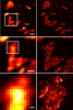
Key to understanding a protein’s biological function is the accurate determination of its spatial distribution inside a cell. Although fluorescent protein markers allow the targeting of specific proteins with molecular precision, much of this information is lost when the resultant fusion proteins are imaged with conventional, diffraction-limited optics. In response, several imaging modalities that are capable of resolution below the diffraction limit (approximately 200 nm) have emerged. Here, both single- and dual-color superresolution imaging of biological structures using photoactivated localization microscopy (PALM) are described. The examples discussed focus on adhesion complexes: dense, protein-filled assemblies that form at the interface between cells and their substrata. A particular emphasis is placed on the instrumentation and photoactivatable fluorescent protein (PA-FP) tags necessary to achieve PALM images at approximately 20 nm resolution in 5 to 30 min in fixed cells.
Commentary: A paper spearheaded by Hari which gives a thorough description of the methods and hardware needed to successfully practice PALM, including cover slip preparation, cell transfection and fixation, drift correction with fiducials, characterization of on/off contrast ratios for different photoactivted fluorescent proteins, identifying PALM-suitable cells, and mechanical and optical components of a PALM system.
A subclass of fluorescent proteins (FPs), large Stokes shift (LSS) FP, are characterized by increased spread between excitation and emission maxima. We report a photoswitchable variant of a red FP with an LSS, PSLSSmKate, which initially exhibits excitation and emission at 445 and 622 nm, but violet irradiation photoswitches PSLSSmKate into a common red form with excitation and emission at 573 and 621 nm. We characterize spectral, photophysical, and biochemical properties of PSLSSmKate in vitro and in mammalian cells and determine its crystal structure in the LSS form. Mass spectrometry, mutagenesis, and spectroscopy of PSLSSmKate allow us to propose molecular mechanisms for the LSS, pH dependence, and light-induced chromophore transformation. We demonstrate the applicability of PSLSSmKate to superresolution photoactivated localization microscopy and protein dynamics in live cells. Given its promising properties, we expect that PSLSSmKate-like phenotype will be further used for photoactivatable imaging and tracking multiple populations of intracellular objects.
Polymorphism is a key feature of amyloid fibril structures but it remains challenging to explain these variations for a particular sample. Here, we report electron cryomicroscopy-based reconstructions from different fibril morphologies formed by a peptide fragment from an amyloidogenic immunoglobulin light chain. The observed fibril morphologies vary in the number and cross-sectional arrangement of a structurally conserved building block. A comparison with the theoretically possible constellations reveals the experimentally observed spectrum of fibril morphologies to be governed by opposing sets of forces that primarily arise from the β-sheet twist, as well as peptide-peptide interactions within the fibril cross-section. Our results provide a framework for rationalizing and predicting the structure and polymorphism of cross-β fibrils, and suggest that a small number of physical parameters control the observed fibril architectures.
Targeting deep brain structures during electrophysiology and injections requires intensive training and expertise. Even with experience, researchers often can't be certain that a probe is placed precisely in a target location and this complexity scales with the number of simultaneous probes used in an experiment. Here, we present Pinpoint, open-source software that allows for interactive exploration of stereotaxic insertion plans. Once an insertion plan is created, Pinpoint allows users to save these online and share them with collaborators. 3D modeling tools allow users to explore their insertions alongside rig and implant hardware and ensure plans are physically possible. Probes in Pinpoint can be linked to electronic micro-manipulators allowing real-time visualization of current brain region targets alongside neural data. In addition, Pinpoint can control manipulators to automate and parallelize the insertion process. Compared to previously available software, Pinpoint's easy access through web browsers, extensive features, and real-time experiment integration enable more efficient and reproducible recordings.
Power-law scaling in coarse-grained data suggests critical dynamics, but the true source of this scaling often remains unclear. Here, we analyze neural activity recorded during spatial navigation, reproducing power-law scaling under a phenomenological renormalization group (PRG) procedure that clusters units by activity similarity. Such scaling was previously linked to criticality. Here, we show that the iterative nature of the procedure itself leads to the emergence of power laws when applied to heterogeneous, non-interacting units obeying spatially structured activity without requiring critical interactions. Furthermore, the scaling exponents produced by heteregeneous non-interacting units match the observed exponents in recorded neural data. A simplified version of the PRG further reveals how heterogeneity smooths transitions across scales, mimicking critical behavior. The resulting exponents depend systematically on system and population size, predictions confirmed by subsampling the data.
Subiculum, the primary efferent pathway of hippocampus, participates in memory for spatial tasks, relapse to drug abuse, and temporal lobe seizures. Subicular pyramidal neurons exhibit low-threshold burst firing driven by a spike afterdepolarization. Here we report that burst firing can be regulated by stimulation of afferent projections to subiculum. Unlike synaptic plasticity, burst plasticity did not require synaptic depolarization, activation of AMPA or NMDA receptors, or action potential firing. Rather, enhancement of burst firing required synergistic activation of group I, subtype 1 metabotropic glutamate receptors (mGluRs) and muscarinic acetylcholine receptors (mAChR). When either of these receptors was blocked, a suppression of bursting was revealed, which in turn was blocked by antagonists of group I, subtype 5 mGluRs. These results indicate that the output of subiculum can be strongly and bidirectionally regulated by activation of glutamatergic inputs within the hippocampus and cholinergic afferents from the medial septum.
Although all sensory circuits ascend to higher brain areas where stimuli are represented in sparse, stimulus-specific activity patterns, relatively little is known about sensory coding on the descending side of neural circuits, as a network converges. In insects, mushroom bodies have been an important model system for studying sparse coding in the olfactory system, where this format is important for accurate memory formation. In Drosophila, it has recently been shown that the 2,000 Kenyon cells of the mushroom body converge onto a population of only 34 mushroom body output neurons (MBONs), which fall into 21 anatomically distinct cell types. Here we provide the first, to our knowledge, comprehensive view of olfactory representations at the fourth layer of the circuit, where we find a clear transition in the principles of sensory coding. We show that MBON tuning curves are highly correlated with one another. This is in sharp contrast to the process of progressive decorrelation of tuning in the earlier layers of the circuit. Instead, at the population level, odour representations are reformatted so that positive and negative correlations arise between representations of different odours. At the single-cell level, we show that uniquely identifiable MBONs display profoundly different tuning across different animals, but that tuning of the same neuron across the two hemispheres of an individual fly was nearly identical. Thus, individualized coordination of tuning arises at this level of the olfactory circuit. Furthermore, we find that this individualization is an active process that requires a learning-related gene, rutabaga. Ultimately, neural circuits have to flexibly map highly stimulus-specific information in sparse layers onto a limited number of different motor outputs. The reformatting of sensory representations we observe here may mark the beginning of this sensory-motor transition in the olfactory system.
AMPA-type receptors (AMPARs) are rapidly inserted into synapses undergoing long-term potentiation (LTP) to increase synaptic transmission, but how AMPAR-containing vesicles are selectively trafficked to these synapses during LTP is not known. Here we developed a strategy to label AMPAR GluA1 subunits expressed from the endogenous loci of rat hippocampal neurons such that the motion of GluA1-containing vesicles in time-lapse sequences can be characterized using single-particle tracking and mathematical modeling. We find that GluA1-containing vesicles are confined and concentrated near sites of stimulation-induced plasticity. We show that confinement is mediated by actin polymerization, which hinders the active transport of GluA1-containing vesicles along the length of the dendritic shaft by modulating the rheological properties of the cytoplasm. Actin polymerization also facilitates myosin-mediated transport of GluA1-containing vesicles to exocytic sites. We conclude that neurons utilize F-actin to increase vesicular GluA1 reservoirs and promote exocytosis proximal to the sites of neuronal activity.
AMPA-type receptors (AMPARs) are rapidly inserted into synapses undergoing plasticity to increase synaptic transmission, but it is not fully understood if and how AMPAR-containing vesicles are selectively trafficked to these synapses. Here, we developed a strategy to label AMPAR GluA1 subunits expressed from their endogenous loci in cultured rat hippocampal neurons and characterized the motion of GluA1-containing vesicles using single-particle tracking and mathematical modeling. We find that GluA1-containing vesicles are confined and concentrated near sites of stimulation-induced structural plasticity. We show that confinement is mediated by actin polymerization, which hinders the active transport of GluA1-containing vesicles along the length of the dendritic shaft by modulating the rheological properties of the cytoplasm. Actin polymerization also facilitates myosin-mediated transport of GluA1-containing vesicles to exocytic sites. We conclude that neurons utilize F-actin to increase vesicular GluA1 reservoirs and promote exocytosis proximal to the sites of synaptic activity.