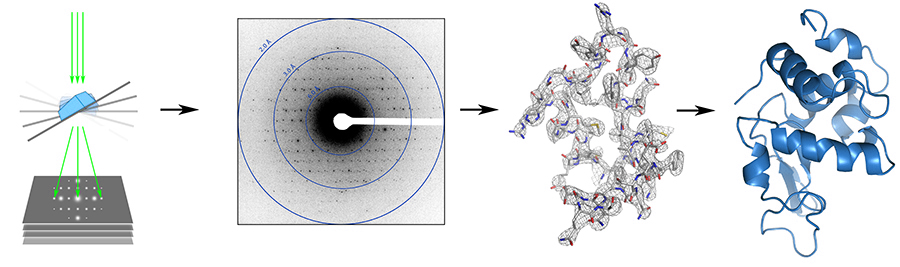
MicroED – Three dimensional electron crystallography of protein microcrystals
Relevant papers
1. Shi D., Nannenga B., Iadanza MG. and Gonen T* (2013). MicroED – Three dimensional electron crystallography of protein microcrystals. eLife – 2:e01345: 1 - 17.
2. Iadanza MG. and Gonen T*. A suite of software for processing MicroED data of extremely small protein crystals. Journal of Applied Crystallography. 47: 1140 – 1145.
3.Nannenga BL, Shi D., Leslie AGW. and Gonen T* (2014). High-resolution structure determination by continuous rotation data collection in MicroED. Nature Methods 11 (9): 927 – 930.
4. Nannenga BL, Shi D., Hattne J., Reyes F. and Gonen T* (2014). Structure of catalase determined by MicroED. eLife 3:e03600: 1 – 11.
5. Hattne J., Reyes FE., Nannenga BL., Shi D., de la Cruz J., Leslie AGW. And Gonen T*. MicroED data collection and processing. Acta Crystallographica section A. A71: 353 - 360 (2015).
6. Rodriguez A.J., Ivanova M., Sawaya MR., Cascio D., Reyes F., Shi D., Sangwan S., Guenther EL., Johnson L, Zhang M., Jiang L., Arbing M., Nannega B., Hattne J., Whitelegge J., Brewster AS., Messerschmidt M., Boutet S., Sauter NK., Gonen T* and Eisenberg D* Structure of the toxic core of a-synuclein from invisible crystals. Nature - In Press (2015).
Relevant Reviews and Book Chapters:
1. Wisedchaisri W., Reichow S.L. and Gonen T*. (2011) Advances in structural and functional analysis of membrane proteins by electron crystallography. Structure. 19:1381-93.2. Wisedchaisri W. and Gonen T*. (2013) Phasing Electron Diffraction Data by Molecular Replacement: Strategy for Structure Determination and Refinement. Methods in Molecular Biology 955: 243 – 272.
3. Gonen T*. (2013) The collection of high-resolution electron diffraction data. Methods in Molecular Biology 955: 153 – 169.
4. Stokes D, Ubarretxena I, Gonen T and Engel A. (2013) High throughout methods in electron crystallography. Methods in Molecular Biology 955: 273 – 296.
5. Nannenga B, Iadanza M, Vollmar B and Gonen T*. (2013) Electron crystallography of membrane proteins: crystallization and screening strategies using negative stain electron microscopy. Current Protocols in Protein Science – 17 (15): 1-11.
6. Nannenga BL. and Gonen T*. Protein structure determination by MicroED. Current Opinion in Structural Biology. 27: 24 - 31 (2014).