Filter
Associated Lab
- Aguilera Castrejon Lab (19) Apply Aguilera Castrejon Lab filter
- Ahrens Lab (73) Apply Ahrens Lab filter
- Aso Lab (42) Apply Aso Lab filter
- Baker Lab (38) Apply Baker Lab filter
- Betzig Lab (116) Apply Betzig Lab filter
- Beyene Lab (15) Apply Beyene Lab filter
- Bock Lab (17) Apply Bock Lab filter
- Branson Lab (55) Apply Branson Lab filter
- Card Lab (43) Apply Card Lab filter
- Cardona Lab (64) Apply Cardona Lab filter
- Chklovskii Lab (13) Apply Chklovskii Lab filter
- Clapham Lab (16) Apply Clapham Lab filter
- Cui Lab (19) Apply Cui Lab filter
- Darshan Lab (12) Apply Darshan Lab filter
- Dennis Lab (3) Apply Dennis Lab filter
- Dickson Lab (46) Apply Dickson Lab filter
- Druckmann Lab (25) Apply Druckmann Lab filter
- Dudman Lab (56) Apply Dudman Lab filter
- Eddy/Rivas Lab (30) Apply Eddy/Rivas Lab filter
- Egnor Lab (11) Apply Egnor Lab filter
- Espinosa Medina Lab (23) Apply Espinosa Medina Lab filter
- Feliciano Lab (12) Apply Feliciano Lab filter
- Fetter Lab (41) Apply Fetter Lab filter
- FIB-SEM Technology (1) Apply FIB-SEM Technology filter
- Fitzgerald Lab (30) Apply Fitzgerald Lab filter
- Freeman Lab (15) Apply Freeman Lab filter
- Funke Lab (46) Apply Funke Lab filter
- Gonen Lab (91) Apply Gonen Lab filter
- Grigorieff Lab (62) Apply Grigorieff Lab filter
- Harris Lab (65) Apply Harris Lab filter
- Heberlein Lab (94) Apply Heberlein Lab filter
- Hermundstad Lab (30) Apply Hermundstad Lab filter
- Hess Lab (80) Apply Hess Lab filter
- Ilanges Lab (4) Apply Ilanges Lab filter
- Jayaraman Lab (48) Apply Jayaraman Lab filter
- Ji Lab (33) Apply Ji Lab filter
- Johnson Lab (6) Apply Johnson Lab filter
- Kainmueller Lab (19) Apply Kainmueller Lab filter
- Karpova Lab (14) Apply Karpova Lab filter
- Keleman Lab (13) Apply Keleman Lab filter
- Keller Lab (76) Apply Keller Lab filter
- Koay Lab (19) Apply Koay Lab filter
- Lavis Lab (158) Apply Lavis Lab filter
- Lee (Albert) Lab (34) Apply Lee (Albert) Lab filter
- Leonardo Lab (23) Apply Leonardo Lab filter
- Li Lab (32) Apply Li Lab filter
- Lippincott-Schwartz Lab (180) Apply Lippincott-Schwartz Lab filter
- Liu (Yin) Lab (8) Apply Liu (Yin) Lab filter
- Liu (Zhe) Lab (65) Apply Liu (Zhe) Lab filter
- Looger Lab (138) Apply Looger Lab filter
- Magee Lab (49) Apply Magee Lab filter
- Menon Lab (18) Apply Menon Lab filter
- Murphy Lab (13) Apply Murphy Lab filter
- O'Shea Lab (8) Apply O'Shea Lab filter
- Otopalik Lab (13) Apply Otopalik Lab filter
- Pachitariu Lab (52) Apply Pachitariu Lab filter
- Pastalkova Lab (19) Apply Pastalkova Lab filter
- Pavlopoulos Lab (19) Apply Pavlopoulos Lab filter
- Pedram Lab (15) Apply Pedram Lab filter
- Podgorski Lab (16) Apply Podgorski Lab filter
- Reiser Lab (55) Apply Reiser Lab filter
- Riddiford Lab (44) Apply Riddiford Lab filter
- Romani Lab (51) Apply Romani Lab filter
- Rubin Lab (149) Apply Rubin Lab filter
- Saalfeld Lab (64) Apply Saalfeld Lab filter
- Satou Lab (18) Apply Satou Lab filter
- Scheffer Lab (38) Apply Scheffer Lab filter
- Schreiter Lab (70) Apply Schreiter Lab filter
- Schulze Lab (1) Apply Schulze Lab filter
- Sgro Lab (23) Apply Sgro Lab filter
- Shroff Lab (31) Apply Shroff Lab filter
- Simpson Lab (23) Apply Simpson Lab filter
- Singer Lab (80) Apply Singer Lab filter
- Spruston Lab (98) Apply Spruston Lab filter
- Stern Lab (160) Apply Stern Lab filter
- Sternson Lab (54) Apply Sternson Lab filter
- Stringer Lab (41) Apply Stringer Lab filter
- Svoboda Lab (136) Apply Svoboda Lab filter
- Tebo Lab (35) Apply Tebo Lab filter
- Tervo Lab (9) Apply Tervo Lab filter
- Tillberg Lab (22) Apply Tillberg Lab filter
- Tjian Lab (64) Apply Tjian Lab filter
- Truman Lab (88) Apply Truman Lab filter
- Turaga Lab (53) Apply Turaga Lab filter
- Turner Lab (38) Apply Turner Lab filter
- Vale Lab (8) Apply Vale Lab filter
- Voigts Lab (4) Apply Voigts Lab filter
- Wang (Meng) Lab (29) Apply Wang (Meng) Lab filter
- Wang (Shaohe) Lab (25) Apply Wang (Shaohe) Lab filter
- Wong-Campos Lab (1) Apply Wong-Campos Lab filter
- Wu Lab (9) Apply Wu Lab filter
- Zlatic Lab (28) Apply Zlatic Lab filter
- Zuker Lab (25) Apply Zuker Lab filter
Associated Project Team
- CellMap (13) Apply CellMap filter
- COSEM (3) Apply COSEM filter
- FIB-SEM Technology (5) Apply FIB-SEM Technology filter
- Fly Descending Interneuron (12) Apply Fly Descending Interneuron filter
- Fly Functional Connectome (14) Apply Fly Functional Connectome filter
- Fly Olympiad (5) Apply Fly Olympiad filter
- FlyEM (56) Apply FlyEM filter
- FlyLight (50) Apply FlyLight filter
- GENIE (47) Apply GENIE filter
- Integrative Imaging (9) Apply Integrative Imaging filter
- Larval Olympiad (2) Apply Larval Olympiad filter
- MouseLight (18) Apply MouseLight filter
- NeuroSeq (1) Apply NeuroSeq filter
- ThalamoSeq (1) Apply ThalamoSeq filter
- Tool Translation Team (T3) (29) Apply Tool Translation Team (T3) filter
- Transcription Imaging (49) Apply Transcription Imaging filter
Publication Date
- 2026 (70) Apply 2026 filter
- 2025 (222) Apply 2025 filter
- 2024 (210) Apply 2024 filter
- 2023 (158) Apply 2023 filter
- 2022 (192) Apply 2022 filter
- 2021 (194) Apply 2021 filter
- 2020 (196) Apply 2020 filter
- 2019 (202) Apply 2019 filter
- 2018 (232) Apply 2018 filter
- 2017 (217) Apply 2017 filter
- 2016 (209) Apply 2016 filter
- 2015 (252) Apply 2015 filter
- 2014 (236) Apply 2014 filter
- 2013 (194) Apply 2013 filter
- 2012 (190) Apply 2012 filter
- 2011 (190) Apply 2011 filter
- 2010 (161) Apply 2010 filter
- 2009 (158) Apply 2009 filter
- 2008 (140) Apply 2008 filter
- 2007 (106) Apply 2007 filter
- 2006 (92) Apply 2006 filter
- 2005 (67) Apply 2005 filter
- 2004 (57) Apply 2004 filter
- 2003 (58) Apply 2003 filter
- 2002 (39) Apply 2002 filter
- 2001 (28) Apply 2001 filter
- 2000 (29) Apply 2000 filter
- 1999 (14) Apply 1999 filter
- 1998 (18) Apply 1998 filter
- 1997 (16) Apply 1997 filter
- 1996 (10) Apply 1996 filter
- 1995 (18) Apply 1995 filter
- 1994 (12) Apply 1994 filter
- 1993 (10) Apply 1993 filter
- 1992 (6) Apply 1992 filter
- 1991 (11) Apply 1991 filter
- 1990 (11) Apply 1990 filter
- 1989 (6) Apply 1989 filter
- 1988 (1) Apply 1988 filter
- 1987 (7) Apply 1987 filter
- 1986 (4) Apply 1986 filter
- 1985 (5) Apply 1985 filter
- 1984 (2) Apply 1984 filter
- 1983 (2) Apply 1983 filter
- 1982 (3) Apply 1982 filter
- 1981 (3) Apply 1981 filter
- 1980 (1) Apply 1980 filter
- 1979 (1) Apply 1979 filter
- 1976 (2) Apply 1976 filter
- 1973 (1) Apply 1973 filter
- 1970 (1) Apply 1970 filter
- 1967 (1) Apply 1967 filter
Type of Publication
4265 Publications
Showing 3291-3300 of 4265 resultsSelective serotonin reuptake inhibitors (SSRIs) are the most prescribed treatment for individuals experiencing major depressive disorder (MDD). The therapeutic mechanisms that take place before, during, or after SSRIs bind the serotonin transporter (SERT) are poorly understood, partially because no studies exist of the cellular and subcellular pharmacokinetic properties of SSRIs in living cells. We studied escitalopram and fluoxetine using new intensity- based drug-sensing fluorescent reporters (“iDrugSnFRs”) targeted to the plasma membrane (PM), cytoplasm, or endoplasmic reticulum (ER) of cultured neurons and mammalian cell lines. We also employed chemical detection of drug within cells and phospholipid membranes. The drugs attain equilibrium in neuronal cytoplasm and ER, at approximately the same concentration as the externally applied solution, with time constants of a few s (escitalopram) or 200-300 s (fluoxetine). Simultaneously, the drugs accumulate within lipid membranes by ≥ 18-fold (escitalopram) or 180-fold (fluoxetine), and possibly by much larger factors. Both drugs leave cytoplasm, lumen, and membranes just as quickly during washout. We synthesized membrane-impermeant quaternary amine derivatives of the two SSRIs. The quaternary derivatives are substantially excluded from membrane, cytoplasm, and ER for > 2.4 h. They inhibit SERT transport-associated currents 6- or 11-fold less potently than the SSRIs (escitalopram or fluoxetine derivative, respectively), providing useful probes for distinguishing compartmentalized SSRI effects. Although our measurements are orders of magnitude faster than the “therapeutic lag” of SSRIs, these data suggest that SSRI-SERT interactions within organelles or membranes may play roles during either the therapeutic effects or the “antidepressant discontinuation syndrome”.
Selective serotonin reuptake inhibitors (SSRIs) are the most prescribed treatment for individuals experiencing major depressive disorder (MDD). The therapeutic mechanisms that take place before, during, or after SSRIs bind the serotonin transporter (SERT) are poorly understood, partially because no studies exist of the cellular and subcellular pharmacokinetic properties of SSRIs in living cells. We studied escitalopram and fluoxetine using new intensity-based drug-sensing fluorescent reporters ("iDrugSnFRs") targeted to the plasma membrane (PM), cytoplasm, or endoplasmic reticulum (ER) of cultured neurons and mammalian cell lines. We also employed chemical detection of drug within cells and phospholipid membranes. The drugs attain equilibrium in neuronal cytoplasm and ER, at approximately the same concentration as the externally applied solution, with time constants of a few s (escitalopram) or 200-300 s (fluoxetine). Simultaneously, the drugs accumulate within lipid membranes by ≥ 18-fold (escitalopram) or 180-fold (fluoxetine), and possibly by much larger factors. Both drugs leave cytoplasm, lumen, and membranes just as quickly during washout. We synthesized membrane-impermeant quaternary amine derivatives of the two SSRIs. The quaternary derivatives are substantially excluded from membrane, cytoplasm, and ER for > 2.4 h. They inhibit SERT transport-associated currents 6- or 11-fold less potently than the SSRIs (escitalopram or fluoxetine derivative, respectively), providing useful probes for distinguishing compartmentalized SSRI effects. Although our measurements are orders of magnitude faster than the "therapeutic lag" of SSRIs, these data suggest that SSRI-SERT interactions within organelles or membranes may play roles during either the therapeutic effects or the "antidepressant discontinuation syndrome".Selective serotonin reuptake inhibitors stabilize mood in several disorders. In general, these drugs bind to the serotonin (5-hydroxytryptamine) transporter (SERT), which clears serotonin from CNS and peripheral tissues. SERT ligands are effective and relatively safe; primary care practitioners often prescribe them. However, they have several side effects and require 2 to 6 weeks of continuous administration until they act effectively. How they work remains perplexing, contrasting with earlier assumptions that the therapeutic mechanism involves SERT inhibition followed by increased extracellular serotonin levels. This study establishes that two SERT ligands, fluoxetine and escitalopram, enter neurons within minutes, while simultaneously accumulating in many membranes. Such knowledge will motivate future research, hopefully revealing where and how SERT ligands "engage" their therapeutic target(s).
Fluorescence microscopy and fluorescent protein (FP)-tagged GLUT4 molecule have been great tools to characterize GLUT4 localization and dynamics inside the cell. However, it was difficult to distinguish GLUT4 storage vesicles (GSVs) from other intracellular compartments containing GLUT4 in live cells. Here, we describe the use of IRAP-pHluorin and total internal reflection fluorescence (TIRF) microscopy to selectively visualize GSVs and Rab proteins that associate with GSVs. This assay is also valuable to further defining GSV identity by unraveling other GSV-associated proteins.
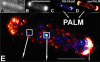
The Escherichia coli chemotaxis network is a model system for biological signal processing. In E. coli, transmembrane receptors responsible for signal transduction assemble into large clusters containing several thousand proteins. These sensory clusters have been observed at cell poles and future division sites. Despite extensive study, it remains unclear how chemotaxis clusters form, what controls cluster size and density, and how the cellular location of clusters is robustly maintained in growing and dividing cells. Here, we use photoactivated localization microscopy (PALM) to map the cellular locations of three proteins central to bacterial chemotaxis (the Tar receptor, CheY, and CheW) with a precision of 15 nm. We find that cluster sizes are approximately exponentially distributed, with no characteristic cluster size. One-third of Tar receptors are part of smaller lateral clusters and not of the large polar clusters. Analysis of the relative cellular locations of 1.1 million individual proteins (from 326 cells) suggests that clusters form via stochastic self-assembly. The super-resolution PALM maps of E. coli receptors support the notion that stochastic self-assembly can create and maintain approximately periodic structures in biological membranes, without direct cytoskeletal involvement or active transport.
Commentary: Our goal as tool developers is to invent methods capable of uncovering new biological insights unobtainable by pre-existing technologies. A terrific example is given by this paper, where grad students Derek Greenfield and Ann McEvoy in Jan Liphardt’s group at Berkeley used our PALM to image the size and position distributions of chemotaxis proteins in E. Coli with unprecedented precision and sensitivity. Their analysis revealed that the cluster sizes follow a stretched exponential distribution, and the density of clusters is highest furthest away from the largest (e.g., polar) clusters. Both observations support a model for passive self-assembly rather than active cytoskeletal assembly of the chemotaxis network.
Cell-free studies have demonstrated how collective action of actin-associated proteins can organize actin filaments into dynamic patterns, such as vortices, asters and stars. Using complementary microscopic techniques, we here show evidence of such self-organization of the actin cortex in living HeLa cells. During cell adhesion, an active multistage process naturally leads to pattern transitions from actin vortices over stars into asters. This process is primarily driven by Arp2/3 complex nucleation, but not by myosin motors, which is in contrast to what has been theoretically predicted and observed in vitro. Concomitant measurements of mechanics and plasma membrane fluidity demonstrate that changes in actin patterning alter membrane architecture but occur functionally independent of macroscopic cortex elasticity. Consequently, tuning the activity of the Arp2/3 complex to alter filament assembly may thus be a mechanism allowing cells to adjust their membrane architecture without affecting their macroscopic mechanical properties.
Reconstructing neuronal circuits at the level of synapses is a central problem in neuroscience, and the focus of the nascent field of connectomics. Previously used to reconstruct the C. elegans wiring diagram, serial-section transmission electron microscopy (ssTEM) is a proven technique for the task. However, to reconstruct more complex circuits, ssTEM will require the automation of image processing. We review progress in the processing of electron microscopy images and, in particular, a semi-automated reconstruction pipeline deployed at Janelia. Drosophila circuits underlying identified behaviors are being reconstructed in the pipeline with the goal of generating a complete Drosophila connectome.
We present semiparametric spectral modeling of the complete larval Drosophila mushroom body connectome. Motivated by a thorough exploratory data analysis of the network via Gaussian mixture modeling (GMM) in the adjacency spectral embedding (ASE) representation space, we introduce the latent structure model (LSM) for network modeling and inference. LSM is a generalization of the stochastic block model (SBM) and a special case of the random dot product graph (RDPG) latent position model, and is amenable to semiparametric GMM in the ASE representation space. The resulting connectome code derived via semiparametric GMM composed with ASE captures latent connectome structure and elucidates biologically relevant neuronal properties.
The GFP-based superecliptic pHluorin (SEP) enables detection of exocytosis and endocytosis, but its performance has not been duplicated in red fluorescent protein scaffolds. Here we describe "semisynthetic" pH-sensitive protein conjugates with organic fluorophores, carbofluorescein, and Virginia Orange that match the properties of SEP. Conjugation to genetically encoded self-labeling tags or antibodies allows visualization of both exocytosis and endocytosis, constituting new bright sensors for these key steps of synaptic transmission.
Fluorescent biosensors are powerful tools for the detection of biochemical events inside cells with high spatiotemporal resolution. Biosensors based on fluorescent proteins often suffer from issues with photostability and brightness. On the other hand, hybrid, chemical–genetic systems present unique opportunities to combine the strengths of synthetic, organic chemistry with biological macromolecules to generate exquisitely tailored semisynthetic sensors.
PURPOSE: To improve the imaging quality of vessel walls with an endoesophageal Wireless Amplified NMR Detector (WAND). METHODS: A cylindrically shaped double-frequency resonator has been constructed with a single metal wire that is self-connected by a pair of nonlinear capacitors. The double-frequency resonator can convert wirelessly provided pumping power into amplified MR signals. This compact design makes the detector easily insertable into a rodent esophagus. RESULTS: The detector has good longitudinal and axial symmetry. Compared to an external surface coil, the WAND can enhance detection sensitivity by at least 5 times, even when the distance separation between the region of interest and the detector's cylindrical surface is twice the detector's own radius. Such detection capability enables us to observe vessel walls near the aortic arch and carotid bifurcation with elevated sensitivity. CONCLUSION: A cylindrical MRI detector integrated with a wireless-powered amplifier has been developed as an endoesophageal detector to enhance detection sensitivity of vessel walls. This detector can greatly improve the imaging quality for vessel regions that are susceptible to atherosclerotic lesions. Magn Reson Med, 2016. © 2016 International Society for Magnetic Resonance in Medicine.