Main Menu (Mobile)- Block
Main Menu - Block
The Janelia Archives
Artifact Name: IsoView Light-Sheet Microscope Science
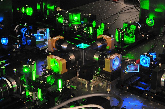
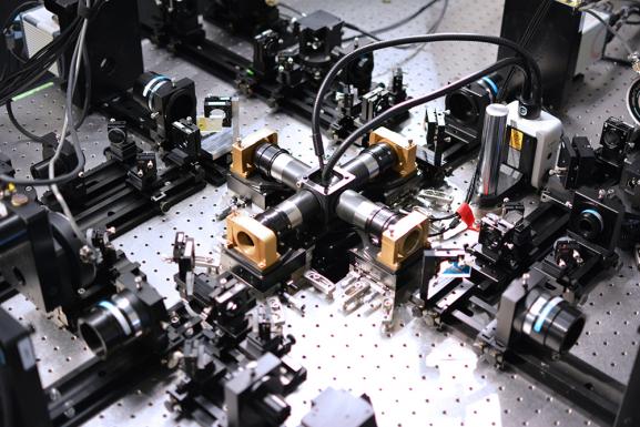
The IsoView light-sheet microscope, developed in the lab of Janelia Group Leader Philipp Keller in 2015, produces images of entire organisms, such as a zebrafish or fruit fly embryo, with enough resolution in all three dimensions that each cell appears as a distinct structure. What's more, it does so within a few hundred milliseconds – fast enough to watch cells migrate as a developing embryo takes shape and to monitor brain activity as neuronal circuits fire.
Previous methods enabled researchers to perform fast three-dimensional imaging of large specimens, but internal cellular structures remained inaccessible. With the IsoView, subcellular structures can be distinguished – without sacrificing speed or field of view. Rather than collecting a single image of a sample with a single objective, the new microscope simultaneously captures images from multiple angles. Each image still suffers from poor resolution along one axis, but combining the most useful data from each image generates a final image with 400 nm resolution in all dimensions – 10-fold better than conventional light-sheet microscopy for large-volume imaging.
The IsoView framework is the result of collaboration between Janelians Raghav Chhetri, an optical physicist who helped develop the image collection methods, and Fernando Amat, a computer scientist who developed algorithms to process the images. The microscope produces approximately 10 terabytes of multiview image data from one hour of imaging, which required developing an entirely new software pipeline to register and deconvolve the images. The 2015 Nature Methods article describing the microscope includes complete building plans for the microscope and associated image-processing software developed by Keller’s team.