MIMMS 2.2 (2024)
Modular In Vivo Multi-photon Microscope 2.2
MIMMS (Modular In vivo Multiphoton Microscopy System) is a modular platform for performing two‐photon laser scanning microscopy (TPLSM) optimized for in vivo applications. The system generally uses commercially available core parts for movement of the objective in the X‐, Y‐, and Z‐axis linear translation and X‐axis rotation for in vivo experiments. The backbone of the design is a movable, raised optical breadboard, providing a large area for affixing optical equipment associated with the microscope. The raised design also provides space for additional testing equipment and allows the entire microscope system to be moved out of the way for 360° access to the working area. The microscope is designed with a modular approach to the components, with interchangeable systems for different laser scanning modalities, moving and fixed objective lens mounting, widefield conventional imaging, and high acceptance, non‐descanned fluorescence detection.
MIMMS 2.2 (2024) Updates:
The Bill of Materials was updated to replace all the components that became obsolete or prohibitively expensive since 2020.
Detection arm module:
- Redesign of the detection arm to accommodate a new piezo based objective scanner from PI (the old one is now obsolete), the piezo objective scanner from Thorlabs, as well as the stationary objective option (no fast-z scanner).
- Complete redesign of the bottom part of the detection arm to make it stiffer and improve stability for the applications where an objective lens needs to be fast scanned through the full stroke at the maximum achievable scan rates.
- ROI RGG laser scanner:
- Complete redesign of the galvo-galvo submodule to accommodate for the ScannerMax galvos. In addition, miniature XY translation stages were added to facilitate precise alignment of the scanned laser beam onto the optical axis of the microscope.
Widefield/1p-epifluorescence and DMD-based opto-stimulation modules:
- Redesigned both to combine them together at the same port. This frees up a port for the second laser scanner module, which is becoming a popular option. In addition, this facilitates an option of widefield imaging with per pixel control of illumination intensity. This can help achieving uniform illumination for imaging of voltage indicators, for example. Refer to MIMMS 2.1 documentation if the DMD option is not needed for the Widefield/1p-epifluorescence module.
Custom Electronics:
- Redesigned the mechanisms for moving the primary dichroic mirror, widefield module mirror and the auxiliary port mirror. Previous design relied on now obsolete components and was expensive.
- Updated Galvo 2U controller CAD model for ScannerMax galvo controller PCB.
- Added CAD models for the PMT controller and the resonant mirror controller.
MIMMS 2.1 (2020) Updates:
- Voice coil-based remote focusing (VC-RF) module is added. This optional module allows for changing the imaging plane position axially without moving the objective lens. The VC-RF unit can prove useful in performing volumetric imaging while maintaining the axial position of the photo-stimulation pattern. In addition, the module can be readily bypassed while maintaining optical alignment through the remaining system. The voice coil offers 6-8 ms step & settle time. This is at least twice faster than piezo-based objective lens scanners.
- DMD-projector-based Illumination module is upgraded with a new projector. The old projector, DLPLIGHTCRAFTER, used in MIMMS 2.0, is now obsolete and can no longer be purchased. The new projector, PRO4500, offers higher resolution, has an all-glass (no plastic optical elements) light engine and 0o optics offset. This significantly simplifies the design and allows higher optical power through the module.
- Bill of Materials is updated and extended to include all components of the MIMMS rig, including all custom-built electronics.
- Documentation, drawings, and pictures are added to help you make your own power supply housings for the dual-galvo and resonant mirror controller PCBs, PMT controller, and PMT preamplifier.
MIMMS 2.0 (2018) Updates:
MIMMS 2.0 (2018) includes various updates for user-friendliness and lowered cost, with more extensive documentation.
- Comprehensive Autodesk Inventor 3D CAD model
- Cost reduction of ~$10,000
- New XYZ stages for improved stability and positional accuracy
- Streamlined components and design features
MIMMS Features always include:
- The design allows for an extensible in vivo 2-photon microscopy system
- The system provides suitable clearance for additional apparatus needed for the specimen.
- MIMMS can be completely moved out of the way to permit full 360° access to the specimen.
- Design created to make customization, modifications, and experimentation possible.
- Assemblies are open and contain more degrees of freedom than monolithic, turn-key systems to allow interoperability with many different key components (lenses, scan systems, etc.) while using well-stocked, commercially available parts, where possible.
Application:
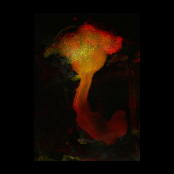
- High-resolution imaging in neurobiology, embryology, and other areas with highly scattering tissues.
- Imaging in highly opaque tissues such as skin.
Opportunity:
For earlier designs, please see MIMMS 1.0 (2016).
Open-source documentation is available for commercial and non-commercial use via the link to Flintbox at the right.
For inquiries, please reference:
Janelia 2018-014

