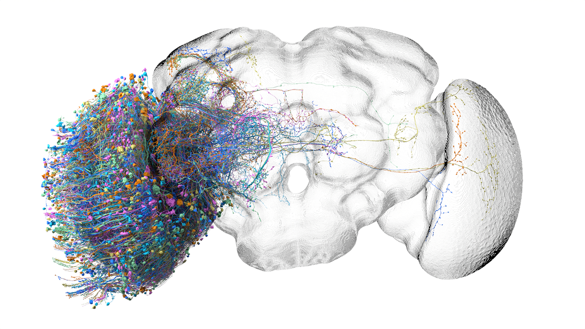
Dipteran flies are highly visual animals and more than half the neurons in the adult brain are found in the optic lobes, the portion of the brain devoted to processing visual information. The anatomy of the optic lobes, characterized by repeating units comprised of neurons with strikingly diverse morphologies, is a beautiful and highly ordered structure that has fascinated neuroanatomists for over 100 years. Here we provide a connectome of the right optic lobe of a male Drosophila melanogaster. This optic lobe is the first proofread and analyzed brain region from an EM volume that contains the entire central nervous system—the central brain, the optic lobes, and the ventral nerve cord, all from the same individual and fully interconnected. The volume contains over 50,000 individual neurons of over 700 distinct types.

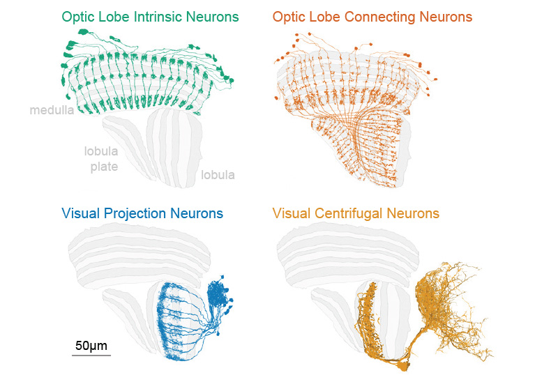
The neurons found in the optic lobe fall into four main groups illustrated above. Optic Lobe Intrinsic Neurons and Optic Lobe Connecting Neurons are confined to the optic lobe, whereas Visual Projection Neurons convey information from the optic lobe to the rest of the brain and Visual Centrifugal Neurons convey information to the optic lobes from the central brain. There are nearly 16,000 intrinsic neurons of nearly 150 different types, 32,000 connecting neurons of over 90 types, 4,500 projection neurons of 350 types and more than 280 centrifugal neurons of more than 100 types.
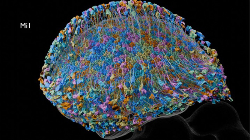
The figure below shows repetitive neurons of a single cell type, Mi1.

Getting started
- Read the paper: Connectome-driven neural inventory of a complete visual system
- Browse the complete catalog of cell types in the Reiser Lab's Cell Type Explorer
- See the Reiser Lab YouTube channel for videos of each cell type and more
- Explore the dataset using neuPrint: an analysis ecosystem for exploring connectomes.
- See the introductory video as well as more detailed documentation.
- Programmatic access via neuprint-python and neuprintr
- View data in neuroglancer:
- https://neuroglancer-demo.appspot.com/#!gs://flyem-optic-lobe/v1.1/optic-lobe-v1.1.json
- EM and segmentation layers are hosted as neuroglancer "precomputed" volumes.
- Downloading subvolumes is possible via TensorStore or cloud-volume
- The complete optic lobe connectome can be downloaded as flat files from this public google bucket.
- If you have questions, problems, and comments, please post on relevant software github pages or in the neuPrint user group.
- The FlyEM optic lobe is licensed under CC-BY.
Videos of Optic Lobe Circuitry
Acknowledgements
Janelia’s FlyEM Project Team acquired and reconstructed the EM dataset in collaboration with the Connectomics group at Google. This work was supported by the Howard Hughes Medical Institute and the Wellcome Trust. We also thank Janelia's FlyLight Project Team, Janelia Connectomics Annotation, Janelia Project Technical Resources, and Janelia's Fly Facility. The Janelia FlyEM Team Project operates under the guidance of its Steering Committee.
Optic Lobe dataset
The Optic Lobe dataset covers the right optic lobe of a male fruit fly. More than half the neurons in the adult fly brain are found in the optic lobes, the portion of the brain devoted to processing visual information.
This image shows the full male fly central nervous system with the right optic lobe highlighted. The medulla is in green, the lobula plate is in purple and the lobula is in yellow-green. Scale bar is 100 µm.
News
- 2024-04-18: Optic Lobe v1.0 released
- 2025-03-10: Optic Lobe v1.1 released
