Filter
Associated Lab
- Aso Lab (2) Apply Aso Lab filter
- Bock Lab (1) Apply Bock Lab filter
- Branson Lab (1) Apply Branson Lab filter
- Card Lab (4) Apply Card Lab filter
- Cardona Lab (1) Apply Cardona Lab filter
- Chklovskii Lab (1) Apply Chklovskii Lab filter
- Dickson Lab (1) Apply Dickson Lab filter
- Fetter Lab (3) Apply Fetter Lab filter
- Funke Lab (4) Apply Funke Lab filter
- Harris Lab (1) Apply Harris Lab filter
- Hess Lab (10) Apply Hess Lab filter
- Jayaraman Lab (3) Apply Jayaraman Lab filter
- Reiser Lab (3) Apply Reiser Lab filter
- Romani Lab (1) Apply Romani Lab filter
- Rubin Lab (12) Apply Rubin Lab filter
- Saalfeld Lab (3) Apply Saalfeld Lab filter
- Scheffer Lab (21) Apply Scheffer Lab filter
- Stern Lab (1) Apply Stern Lab filter
- Truman Lab (1) Apply Truman Lab filter
- Turner Lab (1) Apply Turner Lab filter
Associated Project Team
Publication Date
- 2025 (5) Apply 2025 filter
- 2024 (4) Apply 2024 filter
- 2023 (5) Apply 2023 filter
- 2020 (3) Apply 2020 filter
- 2019 (3) Apply 2019 filter
- 2018 (4) Apply 2018 filter
- 2017 (4) Apply 2017 filter
- 2016 (1) Apply 2016 filter
- 2015 (7) Apply 2015 filter
- 2014 (11) Apply 2014 filter
- 2013 (3) Apply 2013 filter
- 2012 (2) Apply 2012 filter
- 2010 (4) Apply 2010 filter
Type of Publication
56 Publications
Showing 41-50 of 56 resultsThe brain of fruit fly Drosophila melanogaster has been used as a model system for functional analysis of neuronal circuits, including connectomics research, due to its modest size (~700 μm) and availability of abundant molecular genetics tools for visualizing neurons. Three-dimensional (3D) reconstruction of high-resolution images of neurons or circuits visualized with appropriate methods is a critical step for obtaining information such as morphology and connectivity patterns of neuronal circuits. In this chapter, we introduce methods for generating 3D reconstructed images with data acquired from confocal laser scanning microscopy (CLSM) or electron microscopy (EM) to analyze neuronal circuits found in the central nervous system (CNS) of the fruit fly. Comparisons of different algorithms and strategies for reconstructing neuronal circuits, using actual studies as references, will be discussed within this chapter.
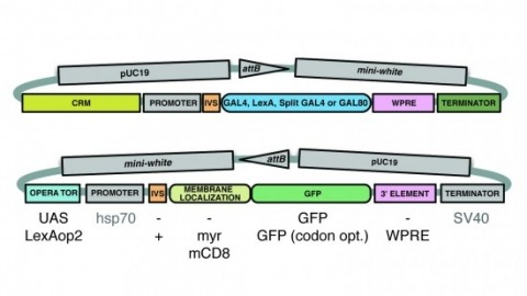
A wide variety of biological experiments rely on the ability to express an exogenous gene in a transgenic animal at a defined level and in a spatially and temporally controlled pattern. We describe major improvements of the methods available for achieving this objective in Drosophila melanogaster. We have systematically varied core promoters, UTRs, operator sequences, and transcriptional activating domains used to direct gene expression with the GAL4, LexA, and Split GAL4 transcription factors and the GAL80 transcriptional repressor. The use of site-specific integration allowed us to make quantitative comparisons between different constructs inserted at the same genomic location. We also characterized a set of PhiC31 integration sites for their ability to support transgene expression of both drivers and responders in the nervous system. The increased strength and reliability of these optimized reagents overcome many of the previous limitations of these methods and will facilitate genetic manipulations of greater complexity and sophistication.
Reconstructing neuronal circuits at the level of synapses is a central problem in neuroscience, and the focus of the nascent field of connectomics. Previously used to reconstruct the C. elegans wiring diagram, serial-section transmission electron microscopy (ssTEM) is a proven technique for the task. However, to reconstruct more complex circuits, ssTEM will require the automation of image processing. We review progress in the processing of electron microscopy images and, in particular, a semi-automated reconstruction pipeline deployed at Janelia. Drosophila circuits underlying identified behaviors are being reconstructed in the pipeline with the goal of generating a complete Drosophila connectome.
Sex differences in behaviour exist across the animal kingdom, typically under strong genetic regulation. In Drosophila, previous work has shown that fruitless and doublesex transcription factors identify neurons driving sexually dimorphic behaviour. However, the organisation of dimorphic neurons into functional circuits remains unclear.We now present the connectome of the entire Drosophila male central nervous system. This contains 166,691 neurons spanning the brain and ventral nerve cord, fully proofread and comprehensively annotated including fruitless and doublesex expression and 11,691 cell types. By comparison with a previous female brain connectome, we provide the first comprehensive description of the differences between male and female brains to synaptic resolution. Of 7,319 cross-matched cell types in the central brain, 114 are dimorphic with an additional 262 male- and 69 female-specific (totalling 4.8% of neurons in males and 2.4% in females).This resource enables analysis of full sensory-to-motor circuits underlying complex behaviours as well as the impact of dimorphic elements. Sex-specific and dimorphic neurons are concentrated in higher brain centres while the sensory and motor periphery are largely isomorphic. Within higher centres, male-specific connections are organised into hotspots defined by male-specific neurons or the presence of male-specific arbours on neurons that are otherwise similar between sexes. Numerous circuit switches reroute sensory information to form conserved, antagonistic circuits controlling opposing behaviours.
Pixel and superpixel classifiers have become essential tools for EM segmentation algorithms. Training these classifiers remains a major bottleneck primarily due to the requirement of completely annotating the dataset which is tedious, error-prone and costly. In this paper, we propose an interactive learning scheme for the superpixel classifier for EM segmentation. Our algorithm is "active semi-supervised" because it requests the labels of a small number of examples from user and applies label propagation technique to generate these queries. Using only a small set (<20%) of all datapoints, the proposed algorithm consistently generates a classifier almost as accurate as that estimated from a complete groundtruth. We provide segmentation results on multiple datasets to show the strength of these classifiers.
Pixel and superpixel classifiers have become essential tools for EM segmentation algorithms. Training these classifiers remains a major bottleneck primarily due to the requirement of completely annotating the dataset which is tedious, error-prone and costly. In this paper, we propose an interactive learning scheme for the superpixel classifier for EM segmentation. Our algorithm is 'active semi-supervised' because it requests the labels of a small number of examples from user and applies label propagation technique to generate these queries. Using only a small set (< 20%) of all datapoints, the proposed algorithm consistently generates a classifier almost as accurate as that estimated from a complete groundtruth. We provide segmentation results on multiple datasets to show the strength of these classifiers.
A central problem in neuroscience is reconstructing neuronal circuits on the synapse level. Due to a wide range of scales in brain architecture such reconstruction requires imaging that is both high-resolution and high-throughput. Existing electron microscopy (EM) techniques possess required resolution in the lateral plane and either high-throughput or high depth resolution but not both. Here, we exploit recent advances in unsupervised learning and signal processing to obtain high depth-resolution EM images computationally without sacrificing throughput. First, we show that the brain tissue can be represented as a sparse linear combination of localized basis functions that are learned using high-resolution datasets. We then develop compressive sensing-inspired techniques that can reconstruct the brain tissue from very few (typically 5) tomographic views of each section. This enables tracing of neuronal processes and, hence, high throughput reconstruction of neural circuits on the level of individual synapses.
We reconstructed the synaptic circuits of seven columns in the second neuropil or medulla behind the fly's compound eye. These neurons embody some of the most stereotyped circuits in one of the most miniaturized of animal brains. The reconstructions allow us, for the first time to our knowledge, to study variations between circuits in the medulla's neighboring columns. This variation in the number of synapses and the types of their synaptic partners has previously been little addressed because methods that visualize multiple circuits have not resolved detailed connections, and existing connectomic studies, which can see such connections, have not so far examined multiple reconstructions of the same circuit. Here, we address the omission by comparing the circuits common to all seven columns to assess variation in their connection strengths and the resultant rates of several different and distinct types of connection error. Error rates reveal that, overall, <1% of contacts are not part of a consensus circuit, and we classify those contacts that supplement (E+) or are missing from it (E-). Autapses, in which the same cell is both presynaptic and postsynaptic at the same synapse, are occasionally seen; two cells in particular, Dm9 and Mi1, form ≥20-fold more autapses than do other neurons. These results delimit the accuracy of developmental events that establish and normally maintain synaptic circuits with such precision, and thereby address the operation of such circuits. They also establish a precedent for error rates that will be required in the new science of connectomics.
Our companion paper (Takemura et al., 2023) introduces the first completely proofread connectome of the nerve cord of an animal that can walk or fly. The base connectome consists of neuronal morphologies and the connections between them. However, in order to efficiently navigate and understand this connectome, it is crucial to have a system of annotations that systematically categorises and names neurons, linking them to the existing literature. In this paper we describe the comprehensive annotation of the VNC connectome, first by a system of hierarchical coarse annotations, then by grouping left-right and serially homologous neurons and eventually by defining systematic cell types for the intrinsic interneurons and sensory neurons of the VNC; descending and motor neurons are typed in (Cheong et al., 2023). We assign a sensory modality to over 5000 sensory neurons, cluster them by connectivity, and identify serially homologous cell types and a layered organisation likely corresponding to peripheral topography. We identify the developmental neuroblast of origin of the large majority of VNC neurons and confirm that (in most cases) all secondary neurons of each hemilineage express a single neurotransmitter. Neuroblast hemilineages are serially repeated along the segments of the nerve cord and generally exhibit consistent hemilineage-to-hemilineage connectivity across neuromeres, supporting the idea that hemilineages are a major organisational feature of the VNC. We also find that more than a third of individual neurons belong to serially homologous cell types, which were crucial for identifying motor neurons and sensory neurons across leg neuropils. Categorising interneurons by their neuropil innervation patterns provides an additional organisation axis. Over half of the intrinsic neurons of the VNC appear dedicated to the legs, with the majority restricted to single leg neuropils; in contrast, inhibitory interneurons connecting different leg neuropils, especially those crossing the midline, appear rarer than anticipated by standard models of locomotor circuitry. Our annotations are being released as part of the neuprint.janelia.org web application and also serve as the basis of programmatic analysis of the connectome through dedicated tools that we describe in this paper.
Analysing computations in neural circuits often uses simplified models because the actual neuronal implementation is not known. For example, a problem in vision, how the eye detects image motion, has long been analysed using Hassenstein-Reichardt (HR) detector or Barlow-Levick (BL) models. These both simulate motion detection well, but the exact neuronal circuits undertaking these tasks remain elusive. We reconstructed a comprehensive connectome of the circuits of Drosophila's motion-sensing T4 cells using a novel EM technique. We uncover complex T4 inputs and reveal that putative excitatory inputs cluster at T4's dendrite shafts, while inhibitory inputs localize to the bases. Consistent with our previous study, we reveal that Mi1 and Tm3 cells provide most synaptic contacts onto T4. We are, however, unable to reproduce the spatial offset between these cells reported previously. Our comprehensive connectome reveals complex circuits that include candidate anatomical substrates for both HR and BL types of motion detectors.