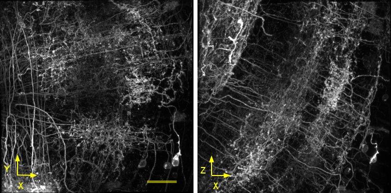
Roughly half the work at Janelia is devoted to “circuit cracking” in Drosophila melanogaster. The Rubin lab has now generated thousands of genetically modified fly lines which allow targeting of single neuron types in the fly brain for imaging or manipulation of activity. Detailed anatomy of particular neurons identifies potential synaptic partners to trace circuits; measurements across animals enable study of plasticity and stereotypy of the circuit.
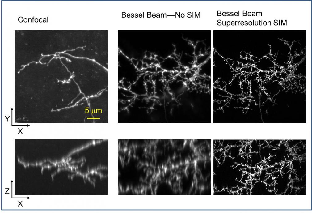
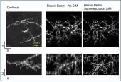
Confocal microscopy is a convenient tool to measure these samples, but lacks the resolution for identifying connections. EM is the gold standard of anatomy measurements, but reconstruction of multiple samples is impractical. Our goal for this technology is to provide high resolution anatomy and connectivity data with the advantages of optical imaging: selective multicolor labeling and sufficient speed to image many samples.
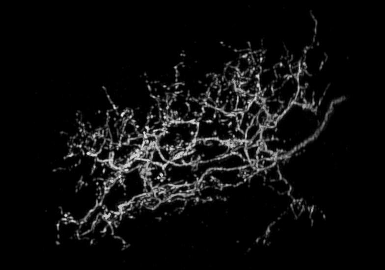
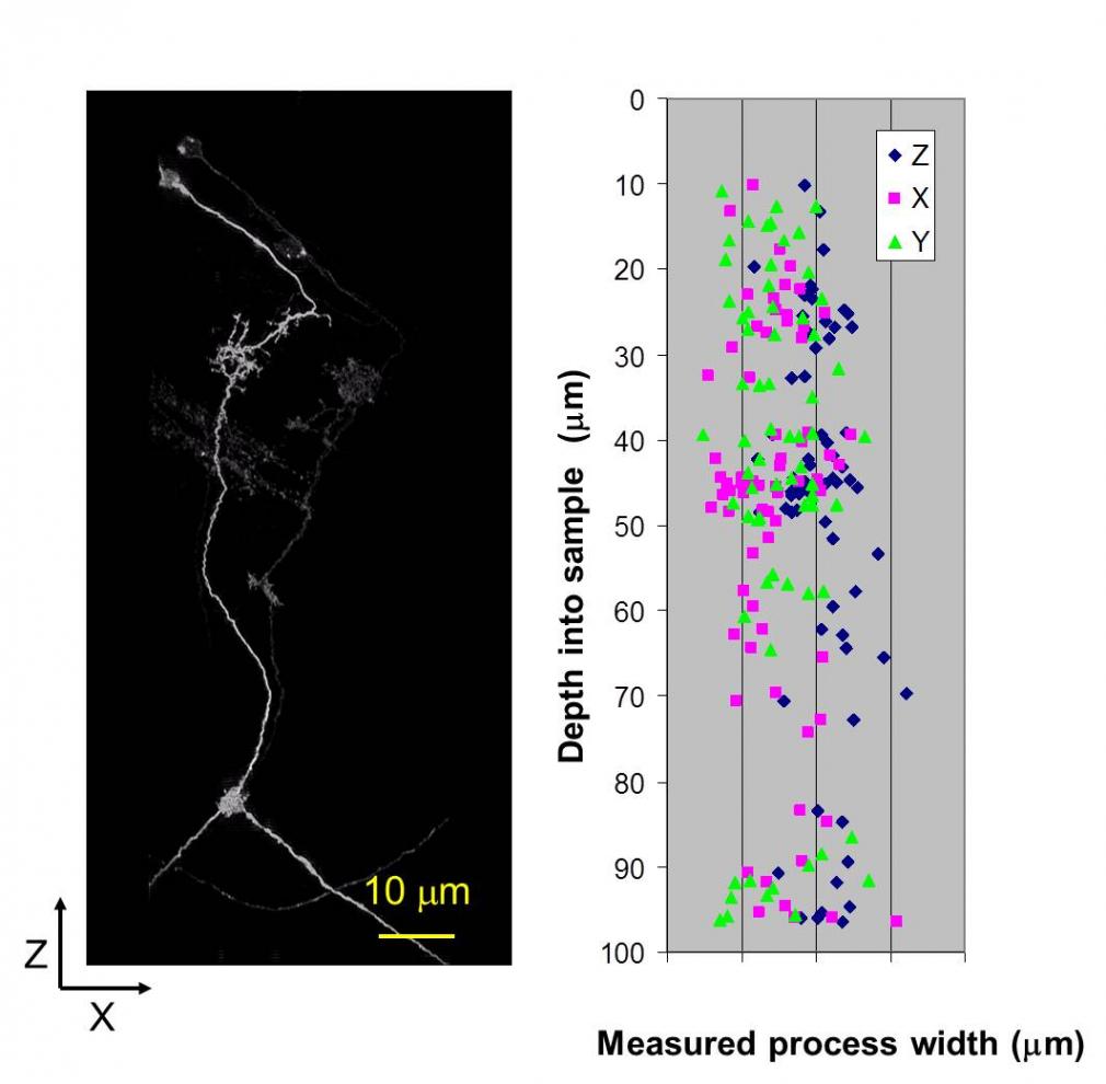
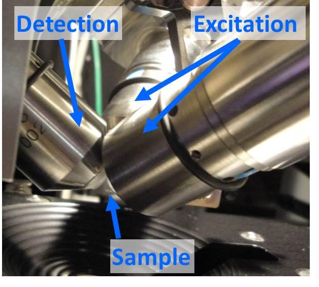
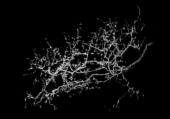
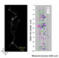
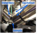
The microscope is designed to image cleared, fluorescently labeled whole Drosophila brains using selective plane illumination microscopy, specifically the Bessel beam super resolution structured illumination techniques pioneered in the Betzig lab. Two excitation objectives and a detection objective positioned along orthogonal axes allow patterning of the light sheet—and thus lateral resolution enhancement—along both x and y of the image plane. Axial resolution enhancement is enabled through the use of very narrow Bessel beams to create the light sheet. We have achieved image resolution of 170 x 170 x 300 nm. Such a voxel size is small enough to reliably infer connectivity from colocalization of fluorescently labeled pre- and post- synaptic markers. Browse the gallery below for sample images.

