Filter
Associated Lab
- Aso Lab (13) Apply Aso Lab filter
- Bock Lab (3) Apply Bock Lab filter
- Branson Lab (3) Apply Branson Lab filter
- Card Lab (7) Apply Card Lab filter
- Cardona Lab (1) Apply Cardona Lab filter
- Dickson Lab (8) Apply Dickson Lab filter
- Funke Lab (1) Apply Funke Lab filter
- Harris Lab (1) Apply Harris Lab filter
- Lavis Lab (1) Apply Lavis Lab filter
- Reiser Lab (7) Apply Reiser Lab filter
- Romani Lab (1) Apply Romani Lab filter
- Rubin Lab (19) Apply Rubin Lab filter
- Saalfeld Lab (2) Apply Saalfeld Lab filter
- Scheffer Lab (1) Apply Scheffer Lab filter
- Simpson Lab (1) Apply Simpson Lab filter
- Singer Lab (1) Apply Singer Lab filter
- Stern Lab (5) Apply Stern Lab filter
- Tillberg Lab (1) Apply Tillberg Lab filter
- Truman Lab (4) Apply Truman Lab filter
- Turner Lab (1) Apply Turner Lab filter
- Zlatic Lab (1) Apply Zlatic Lab filter
Associated Project Team
Publication Date
Type of Publication
48 Publications
Showing 1-10 of 48 resultsFlying insects exhibit remarkable navigational abilities controlled by their compact nervous systems. Optic flow, the pattern of changes in the visual scene induced by locomotion, is a crucial sensory cue for robust self-motion estimation, especially during rapid flight. Neurons that respond to specific, large-field optic flow patterns have been studied for decades, primarily in large flies, such as houseflies, blowflies, and hover flies. The best-known optic-flow sensitive neurons are the large tangential cells of the dipteran lobula plate, whose visual-motion responses, and to a lesser extent, their morphology, have been explored using single-neuron neurophysiology. Most of these studies have focused on the large, Horizontal and Vertical System neurons, yet the lobula plate houses a much larger set of 'optic-flow' sensitive neurons, many of which have been challenging to unambiguously identify or to reliably target for functional studies. Here we report the comprehensive reconstruction and identification of the Lobula Plate Tangential Neurons in an Electron Microscopy (EM) volume of a whole Drosophila brain. This catalog of 58 LPT neurons (per brain hemisphere) contains many neurons that are described here for the first time and provides a basis for systematic investigation of the circuitry linking self-motion to locomotion control. Leveraging computational anatomy methods, we estimated the visual motion receptive fields of these neurons and compared their tuning to the visual consequence of body rotations and translational movements. We also matched these neurons, in most cases on a one-for-one basis, to stochastically labeled cells in genetic driver lines, to the mirror-symmetric neurons in the same EM brain volume, and to neurons in an additional EM data set. Using cell matches across data sets, we analyzed the integration of optic flow patterns by neurons downstream of the LPTs and find that most central brain neurons establish sharper selectivity for global optic flow patterns than their input neurons. Furthermore, we found that self-motion information extracted from optic flow is processed in distinct regions of the central brain, pointing to diverse foci for the generation of visual behaviors.
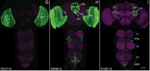
We report the larval CNS expression patterns for 6,650 GAL4 lines based on cis-regulatory regions (CRMs) from the Drosophila genome. Adult CNS expression patterns were previously reported for this collection, thereby providing a unique resource for determining the origins of adult cells. An illustrative example reveals the origin of the astrocyte-like glia of the ventral CNS. Besides larval neurons and glia, the larval CNS contains scattered lineages of immature, adult-specific neurons. Comparison of lineage expression within this large collection of CRMs provides insight into the codes used for designating neuronal types. The CRMs encode both dense and sparse patterns of lineage expression. There is little correlation between brain and thoracic lineages in patterns of sparse expression, but expression in the two regions is highly correlated in the dense mode. The optic lobes, by comparison, appear to use a different set of genetic instructions in their development.
We established a collection of 7,000 transgenic lines of Drosophila melanogaster. Expression of GAL4 in each line is controlled by a different, defined fragment of genomic DNA that serves as a transcriptional enhancer. We used confocal microscopy of dissected nervous systems to determine the expression patterns driven by each fragment in the adult brain and ventral nerve cord. We present image data on 6,650 lines. Using both manual and machine-assisted annotation, we describe the expression patterns in the most useful lines. We illustrate the utility of these data for identifying novel neuronal cell types, revealing brain asymmetry, and describing the nature and extent of neuronal shape stereotypy. The GAL4 lines allow expression of exogenous genes in distinct, small subsets of the adult nervous system. The set of DNA fragments, each driving a documented expression pattern, will facilitate the generation of additional constructs for manipulating neuronal function. synapse was substantially elevated, at the endocytic zone there was no enhanced polymerization activity. We conclude that actin subserves spatially diverse, independently regulated processes throughout spines. Perisynaptic actin forms a uniquely dynamic structure well suited for direct, active regulation of the synapse.
For the overall strategy and methods used to produce the GAL4 lines:
Pfeiffer, B.D., Jenett, A., Hammonds, A.S., Ngo, T.T., Misra, S., Murphy, C., Scully, A., Carlson, J.W., Wan, K.H., Laverty, T.R., Mungall, C., Svirskas, R., Kadonaga, J.T., Doe, C.Q., Eisen, M.B., Celniker, S.E., Rubin, G.M. (2008). Tools for neuroanatomy and neurogenetics in Drosophila. Proc. Natl. Acad. Sci. USA 105, 9715-9720. http://www.pnas.org/content/105/28/9715.full.pdf+html synapse was substantially elevated, at the endocytic zone there was no enhanced polymerization activity. We conclude that actin subserves spatially diverse, independently regulated processes throughout spines. Perisynaptic actin forms a uniquely dynamic structure well suited for direct, active regulation of the synapse.
For data on expression in the embryo:
Manning, L., Purice, M.D., Roberts, J., Pollard, J.L., Bennett, A.L., Kroll, J.R., Dyukareva, A.V., Doan, P.N., Lupton, J.R., Strader, M.E., Tanner, S., Bauer, D., Wilbur, A., Tran, K.D., Laverty, T.R., Pearson, J.C., Crews, S.T., Rubin, G.M., and Doe, C.Q. (2012) Annotated embryonic CNS expression patterns of 5000 GMR GAL4 lines: a resource for manipulating gene expression and analyzing cis-regulatory motifs. Cell Reports (2012) Doi: 10.1016/j.celrep.2012.09.009 http://www.cell.com/cell-reports/fulltext/S2211-1247(12)00290-2 synapse was substantially elevated, at the endocytic zone there was no enhanced polymerization activity. We conclude that actin subserves spatially diverse, independently regulated processes throughout spines. Perisynaptic actin forms a uniquely dynamic structure well suited for direct, active regulation of the synapse.
For data on expression in imaginal discs:
Jory, A., Estella, C., Giorgianni, M.W., Slattery, M., Laverty, T.R., Rubin, G.M., and Mann, R.S. (2012) A survey of 6300 genomic fragments for cis-regulatory activity in the imaginal discs of Drosophila melanogaster. Cell Reports (2012) Doi: 10.1016/j.celrep.2012.09.010 http://www.cell.com/cell-reports/fulltext/S2211-1247(12)00291-4 synapse was substantially elevated, at the endocytic zone there was no enhanced polymerization activity. We conclude that actin subserves spatially diverse, independently regulated processes throughout spines. Perisynaptic actin forms a uniquely dynamic structure well suited for direct, active regulation of the synapse.
For data on expression in the larval nervous system:
Li, H.-H., Kroll, J.R., Lennox, S., Ogundeyi, O., Jeter, J., Depasquale, G., and Truman, J.W. (2013) A GAL4 driver resource for developmental and behavioral studies on the larval CNS of Drosophila. Cell Reports (submitted).
The anatomy of many neural circuits is being characterized with increasing resolution, but their molecular properties remain mostly unknown. Here, we characterize gene expression patterns in distinct neural cell types of the visual system using genetic lines to access individual cell types, the TAPIN-seq method to measure their transcriptomes, and a probabilistic method to interpret these measurements. We used these tools to build a resource of high-resolution transcriptomes for 100 driver lines covering 67 cell types, available at http://www.opticlobe.com. Combining these transcriptomes with recently reported connectomes helps characterize how information is transmitted and processed across a range of scales, from individual synapses to circuit pathways. We describe examples that include identifying neurotransmitters, including cases of apparent co-release, generating functional hypotheses based on receptor expression, as well as identifying strong commonalities between different cell types.
Similar to many insect species, Drosophila melanogaster is capable of maintaining a stable flight trajectory for periods lasting up to several hours. Because aerodynamic torque is roughly proportional to the fifth power of wing length, even small asymmetries in wing size require the maintenance of subtle bilateral differences in flapping motion to maintain a stable path. Flies can even fly straight after losing half of a wing, a feat they accomplish via very large, sustained kinematic changes to both the damaged and intact wings. Thus, the neural network responsible for stable flight must be capable of sustaining fine-scaled control over wing motion across a large dynamic range. In this study, we describe an unusual type of descending neuron (DNg02) that projects directly from visual output regions of the brain to the dorsal flight neuropil of the ventral nerve cord. Unlike many descending neurons, which exist as single bilateral pairs with unique morphology, there is a population of at least 15 DNg02 cell pairs with nearly identical shape. By optogenetically activating different numbers of DNg02 cells, we demonstrate that these neurons regulate wingbeat amplitude over a wide dynamic range via a population code. Using two-photon functional imaging, we show that DNg02 cells are responsive to visual motion during flight in a manner that would make them well suited to continuously regulate bilateral changes in wing kinematics. Collectively, we have identified a critical set of descending neurons that provides the sensitivity and dynamic range required for flight control.
Precise, repeatable genetic access to specific neurons via GAL4/UAS and related methods is a key advantage of Drosophila neuroscience. Neuronal targeting is typically documented using light microscopy of full GAL4 expression patterns, which generally lack the single-cell resolution required for reliable cell type identification. Here we use stochastic GAL4 labeling with the MultiColor FlpOut approach to generate cellular resolution confocal images at large scale. We are releasing aligned images of 74,000 such adult central nervous systems. An anticipated use of this resource is to bridge the gap between neurons identified by electron or light microscopy. Identifying individual neurons that make up each GAL4 expression pattern improves the prediction of split-GAL4 combinations targeting particular neurons. To this end we have made the images searchable on the NeuronBridge website. We demonstrate the potential of NeuronBridge to rapidly and effectively identify neuron matches based on morphology across imaging modalities and datasets.
Techniques that enable precise manipulations of subsets of neurons in the fly central nervous system have greatly facilitated our understanding of the neural basis of behavior. Split-GAL4 driver lines allow specific targeting of cell types in Drosophila melanogaster and other species. We describe here a collection of 3060 lines targeting a range of cell types in the adult Drosophila central nervous system and 1373 lines characterized in third-instar larvae. These tools enable functional, transcriptomic, and proteomic studies based on precise anatomical targeting. NeuronBridge and other search tools relate light microscopy images of these split-GAL4 lines to connectomes reconstructed from electron microscopy images. The collections are the result of screening over 77,000 split hemidriver combinations. In addition to images and fly stocks for these well-characterized lines, we make available 300,000 new 3D images of other split-GAL4 lines.
The fruit fly Drosophila melanogaster is an important model organism for neuroscience with a wide array of genetic tools that enable the mapping of individuals neurons and neural subtypes. Brain templates are essential for comparative biological studies because they enable analyzing many individuals in a common reference space. Several central brain templates exist for Drosophila, but every one is either biased, uses sub-optimal tissue preparation, is imaged at low resolution, or does not account for artifacts. No publicly available Drosophila ventral nerve cord template currently exists. In this work, we created high-resolution templates of the Drosophila brain and ventral nerve cord using the best-available technologies for imaging, artifact correction, stitching, and template construction using groupwise registration. We evaluated our central brain template against the four most competitive, publicly available brain templates and demonstrate that ours enables more accurate registration with fewer local deformations in shorter time.
Descending neurons (DNs) occupy a key position in the sensorimotor hierarchy, conveying signals from the brain to the rest of the body below the neck. In Drosophila melanogaster flies, approximately 480 DN cell types have been described from electron-microscopy image datasets. Genetic access to these cell types is crucial for further investigation of their role in generating behaviour. We previously conducted the first large-scale survey of Drosophila melanogaster DNs, describing 98 unique cell types from light microscopy and generating cell-type-specific split-Gal4 driver lines for 65 of them. Here, we extend our previous work, describing the morphology of 137 additional DN types from light microscopy, bringing the total number DN types identified in light microscopy datasets to 235, or nearly 50%. In addition, we produced 500 new sparse split-Gal4 driver lines and compiled a list of previously published DN lines from the literature for a combined list of 738 split-Gal4 driver lines targeting 171 DN types.
Knowledge of one’s own behavioral state—whether one is walking, grooming, or resting—is critical for contextualizing sensory cues including interpreting visual motion and tracking odor sources. Additionally, awareness of one’s own posture is important to avoid initiating destabilizing or physically impossible actions. Ascending neurons (ANs), interneurons in the vertebrate spinal cord or insect ventral nerve cord (VNC) that project to the brain, may provide such high-fidelity behavioral state signals. However, little is known about what ANs encode and where they convey signals in any brain. To address this gap, we performed a large-scale functional screen of AN movement encoding, brain targeting, and motor system patterning in the adult fly, Drosophila melanogaster. Using a new library of AN sparse driver lines, we measured the functional properties of 247 genetically-identifiable ANs by performing two-photon microscopy recordings of neural activity in tethered, behaving flies. Quantitative, deep network-based neural and behavioral analyses revealed that ANs nearly exclusively encode high-level behaviors—primarily walking as well as resting and grooming—rather than low-level joint or limb movements. ANs that convey self-motion—resting, walking, and responses to gust-like puff stimuli—project to the brain’s anterior ventrolateral protocerebrum (AVLP), a multimodal, integrative sensory hub, while those that encode discrete actions—eye grooming, turning, and proboscis extension—project to the brain’s gnathal ganglion (GNG), a locus for action selection. The structure and polarity of AN projections within the VNC are predictive of their functional encoding and imply that ANs participate in motor computations while also relaying state signals to the brain. Illustrative of this are ANs that temporally integrate proboscis extensions over tens-of-seconds, likely through recurrent interconnectivity. Thus, in line with long-held theoretical predictions, ascending populations convey high-level behavioral state signals almost exclusively to brain regions implicated in sensory feature contextualization and action selection.

